. 2016 Apr 18;82(9):2585-94.
doi: 10.1128/AEM.04070-15. Print 2016 May.

Mechanisms of Bacterial (Serratia marcescens) Attachment to, Migration along, and Killing of Fungal Hyphae

Affiliations

Mechanisms of Bacterial (Serratia marcescens) Attachment to, Migration along, and Killing of Fungal Hyphae

Tal Hover et al. Appl Environ Microbiol. .

Abstract

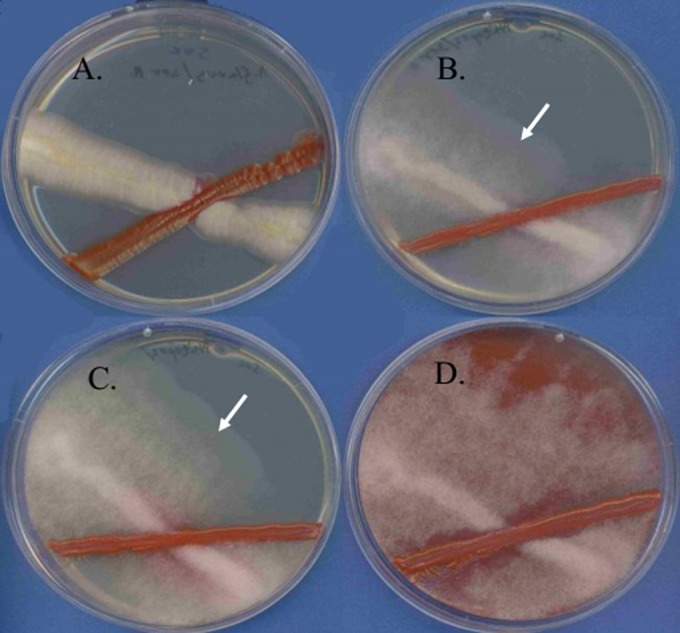
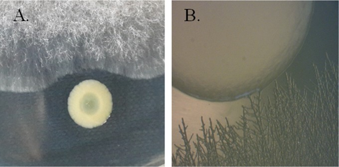
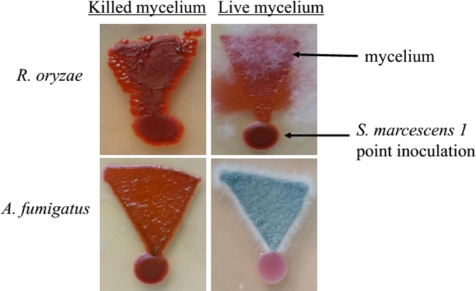
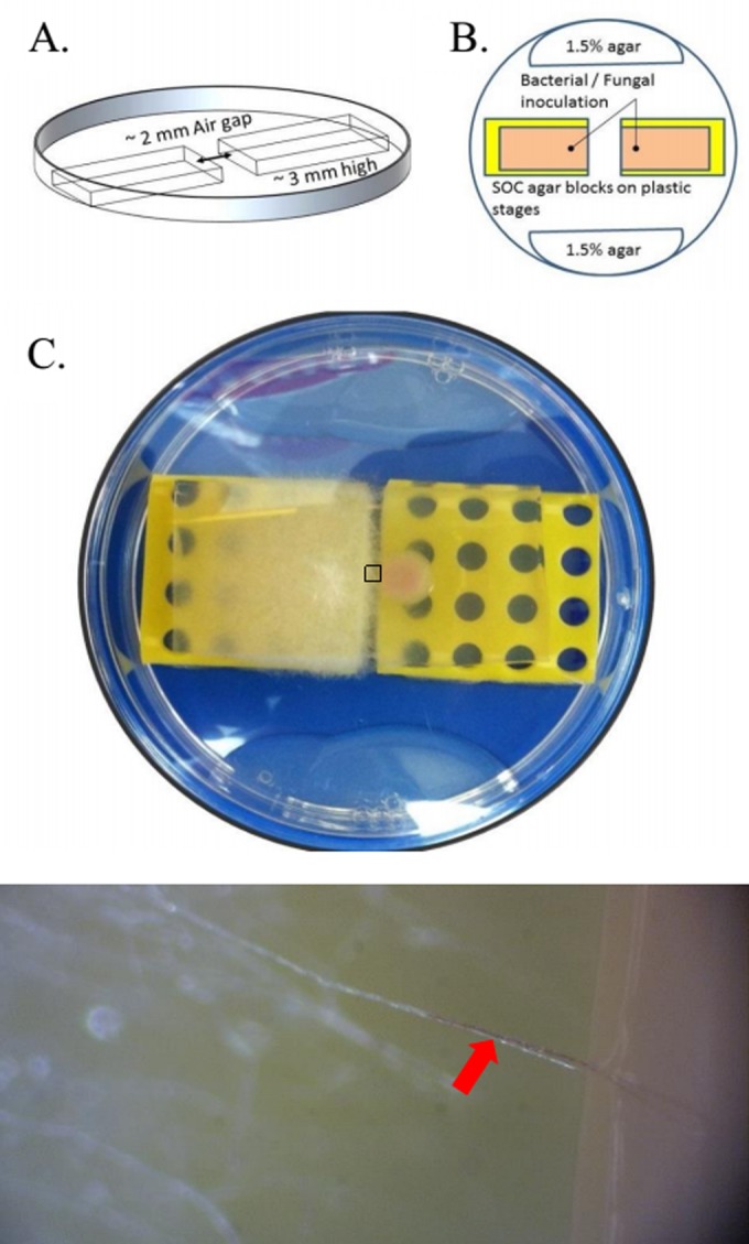
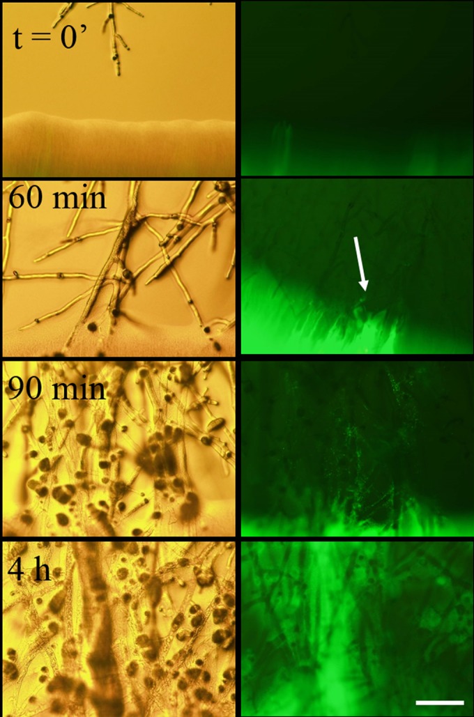
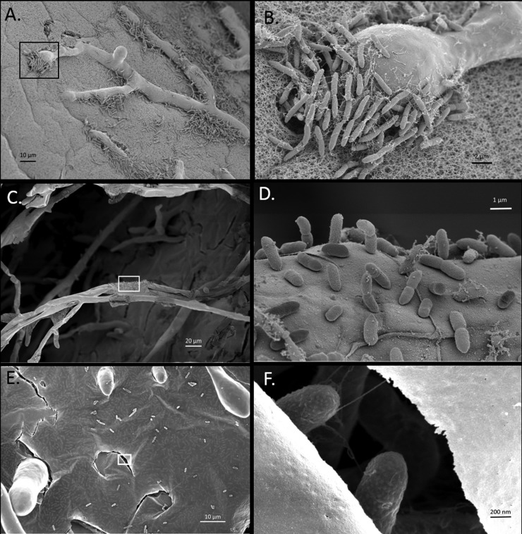
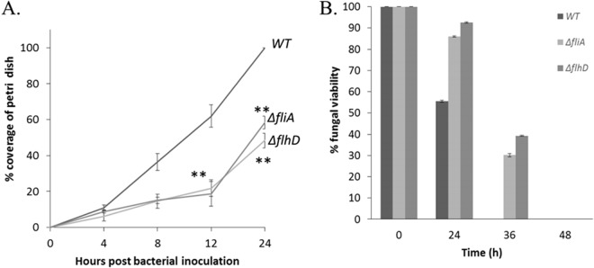
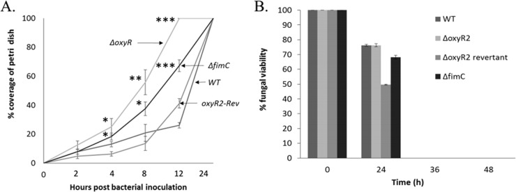
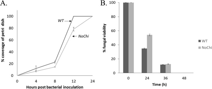
We have found a remarkable capacity for the ubiquitous Gram-negative rod bacterium Serratia marcescens to migrate along and kill the mycelia of zygomycete molds. This migration was restricted to zygomycete molds and several basidiomycete species. No migration was seen on any molds of the phylum Ascomycota. S. marcescens migration did not require fungal viability or surrounding growth medium, as bacteria migrated along aerial hyphae as well.S. marcescens did not exhibit growth tropism toward zygomycete mycelium. Bacterial migration along hyphae proceeded only when the hyphae grew into the bacterial colony. S. marcescens cells initially migrated along the hyphae, forming attached microcolonies that grew and coalesced to generate a biofilm that covered and killed the mycelium. Flagellum-defective strains of S. marcescens were able to migrate along zygomycete hyphae, although they were significantly slower than the wild-type strain and were delayed in fungal killing. Bacterial attachment to the mycelium does not necessitate type 1 fimbrial adhesion, since mutants defective in this adhesin migrated equally well as or faster than the wild-type strain. Killing does not depend on the secretion of S. marcescens chitinases, as mutants in which all three chitinase genes were deleted retained wild-type killing abilities. A better understanding of the mechanisms by which S. marcescens binds to, spreads on, and kills fungal hyphae might serve as an excellent model system for such interactions in general; fungal killing could be employed in agricultural fungal biocontrol.


Figures

FIG 1
FIG 1
S. marcescens spreads over Rhizopus oryzae mold. S. marcescens 1 (red) was cocultured at a 45° angle to various mold species (white). (A) Aspergillus flavus AflI and S. marcescens 1 after 48 h of coincubation. No spreading of S. marcescens over A. flavus mycelium is seen. (B to D) R. oryzae and S. marcescens 1 after 24 h (B), 30 h (C), and 48 h (D) of incubation. Spreading of pigmented S. marcescens over R. oryzae mycelium can be clearly seen preceding the growing hyphal tips at 24 and 30 h (arrows) and covering the entire mycelium after 48 h. All samples were incubated at 37°C on SOC plates.
FIG 2
FIG 2
S. marcescens strain RM66262 does not show chemotaxis toward R. oryzae 5698. Bacterial and fungal strains were point inoculated adjacently. (A) No visible change in bacterial colony morphology is observed, indicating that there is no chemotaxis. (B) S. marcescens migration begins only after hyphae have invaded the bacterial colony. Isolates were grown on SOC at 37°C for 24 h. Identical results were seen with S. marcescens 1 and R. oryzae 3465.
FIG 3
FIG 3
S. marcescens 1 migrates over killed mycelium of R. oryzae 3465 and A. fumigatus but fails to migrate on live A. fumigatus mycelium. Following 36 h of incubation at 30°C on SOC plates, red bacterial coloration is visible on the entire mycelial surface and edges of live or killed R. oryzae mycelium (top). In contrast, S. marcescens spreads on killed A. fumigatus mycelium but fails to do so on live mycelium (bottom).
FIG 4
FIG 4
S. marcescens 1 crosses an aerial bridge of R. oryzae 3465 hyphae. (A and B) Scheme of bridging experiment plate, containing two separate raised SOC agar blocks (orange in panel B) inoculated with bacterial and fungal inoculum separately and incubated in a humid chamber. To eliminate bacterial migration in the water condensate, the SOC agar blocks are placed over raised plastic stages (yellow in panels B and C). Humidity is maintained by pouring 3 ml of a 1.5% agar solution at each side of the plate (colorless in panel B). (C) The air gap is bridged by fungal hyphae after <48 h at 37°C. The box enlarged at the bottom of panel C (top view with a binocular microscope) shows that S. marcescens 1 (red arrow) spreads along R. oryzae 3465 aerial hyphae (red arrow), bridging the gap between the agar blocks after 48 h of incubation at 37°C.
FIG 5
FIG 5
Fluorescence microscopy of S. marcescens interacting with R. oryzae mycelium. S. marcescens 66262/pGFP and R. oryzae 3465 were inoculated adjacently on SOC agar plates and incubated at 37°C until the fungal hyphae came into physical contact with the bacterial colony (time zero [t = 0′]). Light and fluorescent (green fluorescent protein [GFP]) microscopy were performed on the contact region at different time points. The arrow indicates initial early colonization of hyphae by S. marcescens bacteria after 60 min of interaction. Scale bar = 50 μm.
FIG 6
FIG 6
High-resolution SEM imaging of S. marcescens interacting with R. oryzae mycelium. S. marcescens RM66262 forms discrete colonies on R. oryzae 3465 hyphae during early (1 to 2 h) contact on the surface of SOC agar plates (A; the inset in panel A is enlarged in panel B), on aerial hyphae (C; the inset in panel C is enlarged in panel D), later (4 h) forming a mature biofilm (E; the inset in panel E is enlarged in panel F) covering the hyphae. (F) Note the numerous bacteria visible just below the surface of the biofilm in panel E that can be clearly seen where the biofilm surface is disrupted.
FIG 7
FIG 7
S. marcescens mutants lacking a flagellum show impaired migration and killing of R. oryzae 3465. (A) To measure bacterial spreading, the S. marcescens RM66262 wild type (WT), ΔflhD mutant (defective in the flagellar master regulator), and ΔfliA mutant (defective in the flagellar class 2 gene) were inoculated separately in the center of SOC agar petri dishes coated with 12-h-old R. oryzae 3465 mycelia and incubated at 30°C. Bacterial migration was measured by periodic replication of the plates onto fresh SOC medium. The number of new bacterial colonies was used as a marker of bacterial migration rate. (B) To measure hyphal killing, S. marcescens strains were inoculated in the center of 1.5-cm SOC wells coated with 12-h-old R. oryzae 3465 mycelia and incubated at 30°C. Hyphal killing was measured by removing the agar plugs from the wells, incubating them on SOC-kanamycin plates, and scoring the plates for fungal growth, as described in Materials and Methods. The data are representative of the results from three experiments; shown are averages ± standard deviations (SD) from triplicate samples. **, P < 0.005 between mutant and WT strains.
FIG 8
FIG 8
Fimbria-defective S. marcescens mutants migrate faster than the wild type along R. oryzae hyphae. (A) Migration along a preformed R. oryzae 3465 mycelium by the S. marcescens CMS 376 wild type, ΔoxyR2 mutant (defective in the oxidative-stress reaction and fimbrial expression), and ΔfimC mutant (defective in fimbrial structural genes). Strains were inoculated separately on the center of an R. oryzae 3465 mycelium and incubated at 30°C. Bacterial migration was measured, as described in Materials and Methods. (B) Killing of hyphae by S. marcescens fimbria-defective mutants is unchanged relative to that of the CMS 376 wild-type strain. The data are representative of the results from three experiments; shown are averages ± SD from triplicate samples. *, P < 0. 05; **, P < 0.005; ***, P < 0.0005 between mutant and WT strains.
FIG 9
FIG 9
S. marcescens migration and killing of R. oryzae 3465 do not depend on its ability to secrete chitinases. (A) To measure bacterial spreading, the S. marcescens Db10 wild type and Db10-NoChi strain (lacking detectable secreted chitinase activity) were inoculated separately in the center of SOC agar petri dishes coated with 12-h-old R. oryzae 3465 mycelia and incubated at 30°C. Bacterial migration was measured by periodic replication of the plates onto fresh SOC medium. The number of new bacterial colonies was used as a marker of the bacterial migration rate. (B) To measure hyphal killing, S. marcescens strains were inoculated in the center of 1.5-cm SOC wells coated with 12-h-old R. oryzae 3465 mycelia and incubated at 30°C. Hyphal killing was measured by removing the agar plugs from the wells, incubating them on SOC-kanamycin plates, and scoring the plates for fungal growth, as described in Materials and Methods. The data are representative of the results from three experiments showing the averages ± SD from triplicate samples.

Similar articles

Cited by

References

    1. Furuno S, Pazolt K, Rabe C, Neu TR, Harms H, Wick LY. 2010. Fungal mycelia allow chemotactic dispersal of polycyclic aromatic hydrocarbon-degrading bacteria in water-unsaturated systems. Environ Microbiol 12:1391–1398. - PubMed
    1. Kohlmeier S, Smits TH, Ford RM, Keel C, Harms H, Wick LY. 2005. Taking the fungal highway: mobilization of pollutant-degrading bacteria by fungi. Environ Sci Technol 39:4640–4646. doi:10.1021/es047979z. - DOI - PubMed
    1. Wick LY, Remer R, Wurz B, Reichenbach J, Braun S, Schafer F, Harms H. 2007. Effect of fungal hyphae on the access of bacteria to phenanthrene in soil. Environ Sci Technol 41:500–505. doi:10.1021/es061407s. - DOI - PubMed
    1. Peleg AY, Hogan DA, Mylonakis E. 2010. Medically important bacterial-fungal interactions. Nat Rev Microbiol 8:340–349. doi:10.1038/nrmicro2313. - DOI - PubMed
    1. Hogan DA, Kolter R. 2002. Pseudomonas-Candida interactions: an ecological role for virulence factors. Science 296:2229–2232. doi:10.1126/science.1070784. - DOI - PubMed

Publication types

MeSH terms

LinkOut - more resources