. 2016 Sep 15;33(18):1696-708.
doi: 10.1089/neu.2015.4111. Epub 2016 Apr 8.

Tamoxifen Administration Immediately or 24 Hours after Spinal Cord Injury Improves Locomotor Recovery and Reduces Secondary Damage in Female Rats

Affiliations

Tamoxifen Administration Immediately or 24 Hours after Spinal Cord Injury Improves Locomotor Recovery and Reduces Secondary Damage in Female Rats

Jennifer M Colón et al. J Neurotrauma. .

Abstract

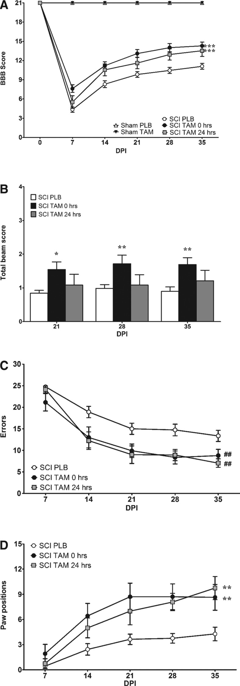
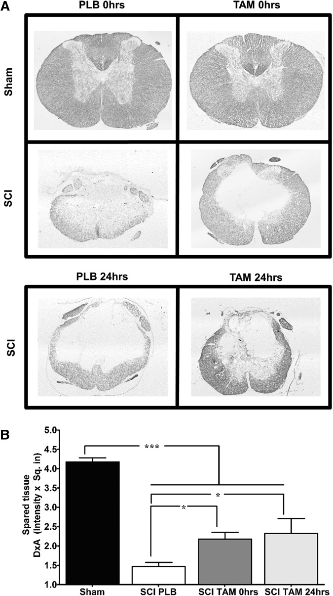
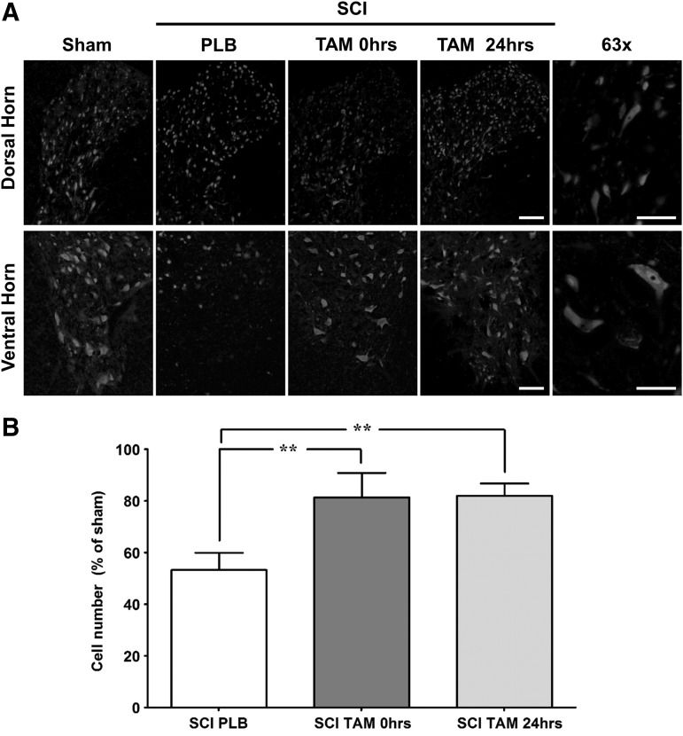
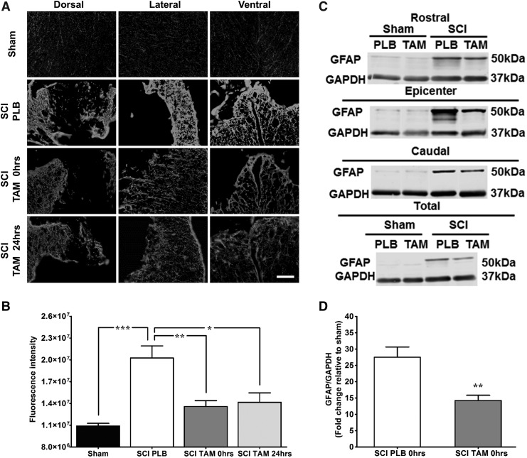
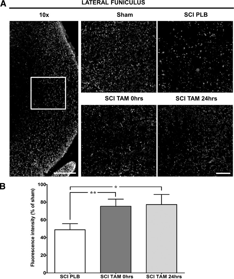
Spinal cord injury (SCI) is a condition with no available cure. The initial physical impact triggers a cascade of molecular and cellular events that generate a nonpermissive environment for cell survival and axonal regeneration. Spinal cord injured patients often arrive at the clinic hours after the initial insult. This indicates the need to study and develop treatments with a long therapeutic window of action and multiactive properties, which target the complex set of events that arise after the initial trauma. We provide evidence that tamoxifen (TAM), a drug approved by the Food and Drug Administration, exerts neuroprotective effects in an animal model when applied up-to 24 h after SCI. We hypothesized that continuous TAM administration will improve functional locomotor recovery by favoring myelin preservation and reducing secondary damage after SCI. Adult female Sprague-Dawley rats (∼230 g) received a moderate contusion to the thoracic (T9-T10) spinal cord, using the MASCIS impactor device. To determine the therapeutic window available for TAM treatment, rats were implanted with TAM pellets (15 mg) immediately or 24 h after SCI. Locomotor function (Basso, Beattie, Bresnahan open field test, grid walk, and beam crossing tests) was assessed weekly for 35 days post-injury. TAM-treated rats showed significant functional locomotor recovery and improved fine movements when treated immediately or 24 h after SCI. Further, TAM increased white matter preservation and reduced secondary damage caused by astrogliosis, axonal degeneration, and cell death after trauma. These results provide evidence for TAM as a potential therapeutic agent to treat SCI up to 24 h after the trauma.

Keywords: astrogliosis; neuroprotection; selective estrogen receptor modulator (SERM); spare tissue; therapeutic drug.


Conflict of interest statement

Author Disclosure Statement No competing financial interests exist.

Figures

<b>FIG. 1.</b>
FIG. 1.
Tamoxifen (TAM) administration immediately and 24 h after spinal cord injury (SCI) improves locomotor recovery and coordination. (A) Sham animals obtained 21 points in the Basso, Beattie, Bresnahan (BBB) open field test at all test points evaluated (0–35 DPI). TAM treatment 0 and 24 h after SCI improved locomotor recovery after SCI (***p < 0.001 treatment, DPI, and interaction). (B) Rats treated with TAM (0 h) showed a significant improvement when crossing the narrow beam during chronic stages 21 (*p < 0.05), 28 DPI, and 35 DPI (**p < 0.01) (treatment, DPI, and interaction). (C,D) TAM improves coordination by reducing the number of errors (##p < 0.01; treatment) and increasing the number of correct paw positions in the grid walk test (**p < 0.01; treatment, DPI, and interaction). Data are the mean ± SEM (sham PLB n = 9, sham TAM n = 9, SCI PLB (rats from t = 0 + 24 h) n = 29, SCI TAM 0 h n = 21, and SCI TAM 24 h n = 12).
<b>FIG. 2.</b>
FIG. 2.
Delayed tamoxifen (TAM) therapy 24 h after spinal cord injury (SCI) increases the amount of white matter spared tissue at the lesion epicenter. (A) Representative transverse spinal cord sections evaluated for Luxol fast blue staining at the lesion epicenter of sham and injured animals at 35 days post-injury (10x magnification). (B) Densitometry analysis of transverse sections at the lesion epicenter revealed that TAM treatment favors white matter preservation when administered immediately and 24 h after SCI. Three sections per animal were evaluated, one-way analysis of variance data are mean ± SEM. Sham (PLB+TAM) n = 13, SCI PLB (rats from t = 0 h + 24 h) n = 6, SCI TAM 0 h n = 7, SCI TAM 24 h n = 3 (*p < 0.05, **p < 0.01, ***p < 0.001).
<b>FIG. 3.</b>
FIG. 3.
Tamoxifen (TAM) promotes neuronal survival in the dorsal and ventral horn at the rostral penumbra after spinal cord injury (SCI). (A) Representative images of NeunN immunoreactivity from the DH or VH in the rostral penumbra at 35 days post-injury (magnification = 20x, scale bar = 100 μm). High power magnification (63x) of dorsal and ventral horn neurons from sham animals (scale bar = 50 μm). (B) Neuronal cell count shows that SCI reduces the total number of neurons in the rostral area. TAM treatment at 0 and 24 h after SCI significantly increases the number of neurons from the DH and VH of injured animals. One-way analysis of variance Tukey's multiple comparison test (**p < 0.01), data are mean ± SEM, sham (PLB+TAM) n = 9, SCI PLB (rats from t = 0 h + 24 h) n = 4, SCI TAM 0 h n = 3, SCI TAM 24 h n = 3.
<b>FIG. 4.</b>
FIG. 4.
Immediate and delayed tamoxifen (TAM) administration after spinal cord injury (SCI) decreased secondary damage caused by astrogliosis. (A) Representative images of glial fibrillary acidic protein (GFAP) immunoreactivity at the lesion epicenter 35 DPI (magnification 20x, scale bar = 100 μm). (B) Relative fluorescence intensity quantification of GFAP immunoreactivity from the epicenter. Sections were divided into anatomical quadrants (dorsal, lateral, and ventral), and the total area was quantified and averaged for the analysis (scale bar = 100 μm). Animals with SCI showed significant increased GFAP immunoreactivity. TAM treatment at 0 and 24 h after SCI reduced secondary damage associated to reactive gliosis at the lesion epicenter according to one-way analysis of variance Tukey multiple comparison test (***p < 0.001, **p < 0.01, *p < 0.05). Data are mean ± SEM, sham (PLB+TAM) n = 11, SCI PLB (rats from t = 0 + 24 h) n = 7, SCI TAM 0 h n = 5, SCI TAM 24 h n = 3. (C) Representative immunoblots for GFAP expression at the rostral, epicenter, caudal levels. Equal amounts of proteins from each area were loaded (12.5 μg total), which represent that TAM treatment reduced astrogliosis extent of damage after SCI. (D) Semi-quantitative analysis revealed that TAM significantly reduced GFAP levels when administered immediately after SCI. Data are mean ± SEM standardized against sham and analyzed by unpaired t test (**p < 0.01). Sham PLB n = 2, sham TAM n = 3, SCI PLB (rats from t = 0 + 24 h) n = 4, SCI TAM n = 6.
<b>FIG. 5.</b>
FIG. 5.
Tamoxifen (TAM) favors axonal preservation at the lateral funiculus in the caudal spinal cord. (A) Representation of the lateral funiculus area photographed and quantified for the analysis (magnification = 10x, scale bar = 200 μm). Representative images of NF-H immunoreactivity, approximately 2 mm caudal to the lesion epicenter at 35 DPI (magnification = 40x, scale bar = 50 μm). (B) Relative fluorescence intensity quantification of NF-H immunoreactivity shows TAM treatment 0 and 24 h after SCI increased the amount of neurofilaments at the lateral funiculus after SCI. Isotype controls were kept for every experiment; IgG1 immunoreactivity was used to calibrate the microscope and quantified to determine the background level of fluorescence. Left and right lateral funiculus were analyzed, and the average of the total quantification was evaluated for statistical significance. One-way analysis of variance Tukey's multiple comparison test (**p < 0.01, *p < 0.05), data are mean ± SEM, sham (PLB+TAM) n = 10, SCI PLB (rats from t = 0 + 24 h) n = 6, SCI TAM 0 h n = 5, SCI TAM 24 h n = 3.

References

    1. Rabchevsky A.G., Patel S.P., and Springer J.E. (2011). Pharmacological interventions for spinal cord injury: where do we stand? How might we step forward? Pharmacol. Ther. 132, 15–29 - PubMed
    1. McKerracher L., and Higuchi H. (2006). Targeting Rho to stimulate repair after spinal cord injury. J. Neurotrauma 23, 309–17 - PubMed
    1. Fitch M.T., and Silver J. (2008). CNS injury, glial scars, and inflammation: Inhibitory extracellular matrices and regeneration failure. Exp. Neurol. 209, 294–301 - PMC - PubMed
    1. Hulsebosch C.E. (2002). Recent advances in pathophysiology and treatment of spinal cord injury. Adv. Physiol. Educ. 26, 238–255 - PubMed
    1. Sofroniew M.V. (2009). Molecular dissection of reactive astrogliosis and glial scar formation. Trends Neurosci. 32, 638–647 - PMC - PubMed

Publication types