

Studying Z-DNA and B- to Z-DNA transitions using a cytosine analogue FRET-pair
- PMID: 26896804
- PMCID: PMC4914084
- DOI: 10.1093/nar/gkw114
Studying Z-DNA and B- to Z-DNA transitions using a cytosine analogue FRET-pair
Abstract
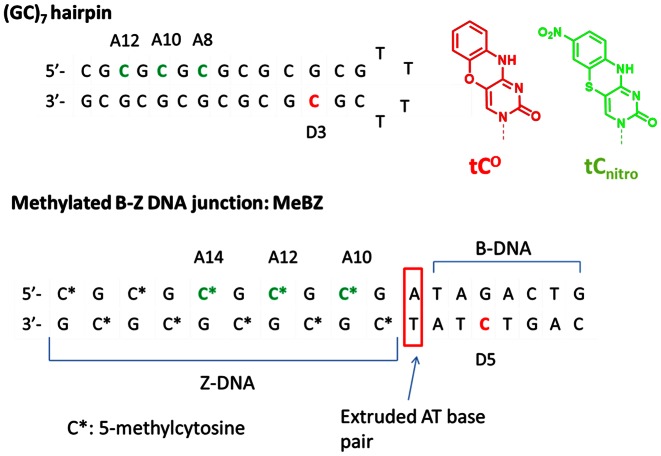
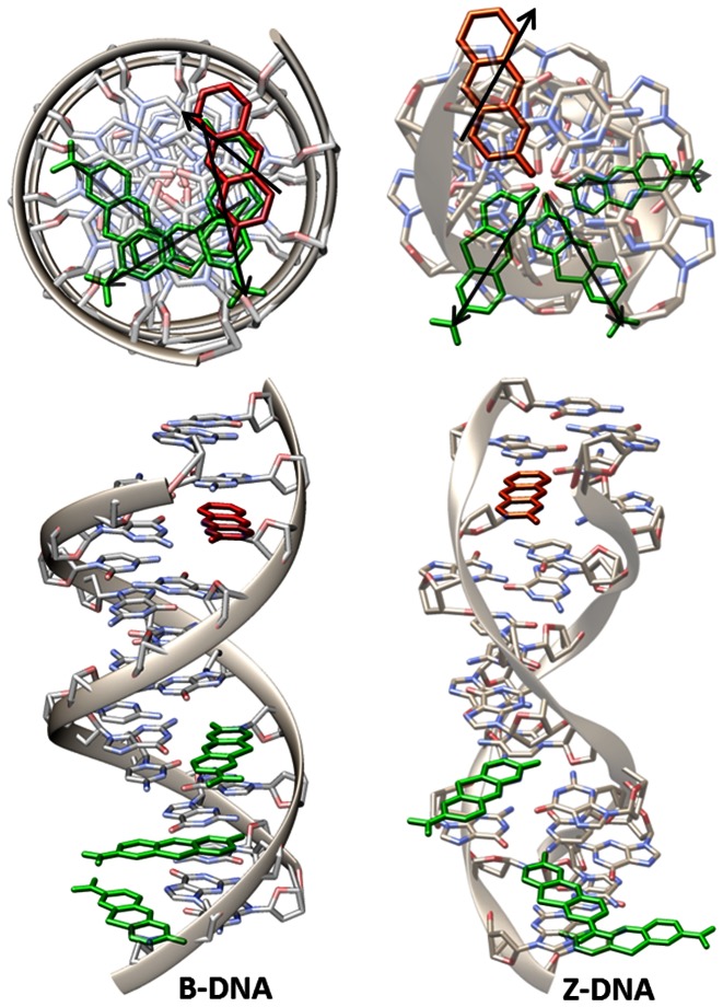
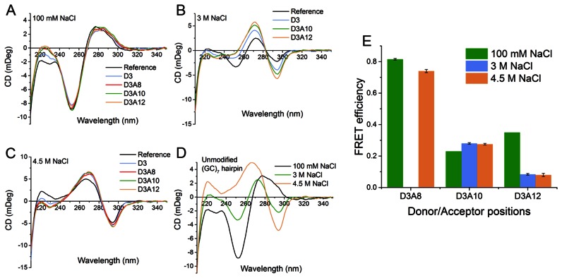
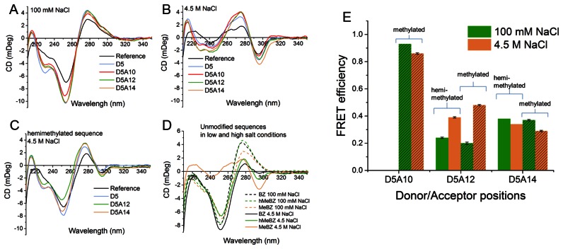
Herein, we report on the use of a tricyclic cytosine FRET pair, incorporated into DNA with different base pair separations, to study Z-DNA and B-Z DNA junctions. With its position inside the DNA structure, the FRET pair responds to a B- to Z-DNA transition with a distinct change in FRET efficiency for each donor/acceptor configuration allowing reliable structural probing. Moreover, we show how fluorescence spectroscopy and our cytosine analogues can be used to determine rate constants for the B- to Z-DNA transition mechanism. The modified cytosines have little influence on the transition and the FRET pair is thus an easily implemented and virtually non-perturbing fluorescence tool to study Z-DNA. This nucleobase analogue FRET pair represents a valuable addition to the limited number of fluorescence methods available to study Z-DNA and we suggest it will facilitate, for example, deciphering the B- to Z-DNA transition mechanism and investigating the interaction of DNA with Z-DNA binding proteins.
© The Author(s) 2016. Published by Oxford University Press on behalf of Nucleic Acids Research.
Figures
Similar articles
-
Fluorescent nucleobase analogues for base-base FRET in nucleic acids: synthesis, photophysics and applications.Beilstein J Org Chem. 2018 Jan 10;14:114-129. doi: 10.3762/bjoc.14.7. eCollection 2018. Beilstein J Org Chem. 2018. PMID: 29441135 Free PMC article. Review.
-
Interbase FRET in RNA: from A to Z.Nucleic Acids Res. 2019 Nov 4;47(19):9990-9997. doi: 10.1093/nar/gkz812. Nucleic Acids Res. 2019. PMID: 31544922 Free PMC article.
-
Characterization of nucleobase analogue FRET acceptor tCnitro.J Phys Chem B. 2010 Jan 21;114(2):1050-6. doi: 10.1021/jp909471b. J Phys Chem B. 2010. PMID: 20039634
-
Nucleic acid base analog FRET-pair facilitating detailed structural measurements in nucleic acid containing systems.J Am Chem Soc. 2009 Apr 1;131(12):4288-93. doi: 10.1021/ja806944w. J Am Chem Soc. 2009. PMID: 19317504
-
Comparative review on left-handed Z-DNA.Front Biosci (Landmark Ed). 2021 Apr 30;26(5):29-35. doi: 10.52586/4922. Front Biosci (Landmark Ed). 2021. PMID: 34027648 Review.
Cited by
-
Fluorescent nucleobase analogues for base-base FRET in nucleic acids: synthesis, photophysics and applications.Beilstein J Org Chem. 2018 Jan 10;14:114-129. doi: 10.3762/bjoc.14.7. eCollection 2018. Beilstein J Org Chem. 2018. PMID: 29441135 Free PMC article. Review.
-
Low dose dimethyl sulfoxide driven gross molecular changes have the potential to interfere with various cellular processes.Sci Rep. 2018 Oct 4;8(1):14828. doi: 10.1038/s41598-018-33234-z. Sci Rep. 2018. PMID: 30287873 Free PMC article.
-
Interbase FRET in RNA: from A to Z.Nucleic Acids Res. 2019 Nov 4;47(19):9990-9997. doi: 10.1093/nar/gkz812. Nucleic Acids Res. 2019. PMID: 31544922 Free PMC article.
-
Autoimmunity and SLE: Factual and Semantic Evidence-Based Critical Analyses of Definitions, Etiology, and Pathogenesis.Front Immunol. 2020 Oct 6;11:569234. doi: 10.3389/fimmu.2020.569234. eCollection 2020. Front Immunol. 2020. PMID: 33123142 Free PMC article. Review.
-
Fluorescent RNA cytosine analogue - an internal probe for detailed structure and dynamics investigations.Sci Rep. 2017 May 24;7(1):2393. doi: 10.1038/s41598-017-02453-1. Sci Rep. 2017. PMID: 28539582 Free PMC article.
References
-
- Choi J., Majima T. Conformational changes of non-B DNA. Chem. Soc. Rev. 2011;40:5893–5909. - PubMed
-
- Belotserkovskii B.P., Mirkin S.M., Hanawalt P.C. DNA sequences that interfere with transcription: Implications for genome function and stability. Chem. Rev. 2013;113:8620–8637. - PubMed
-
- Doluca O., Withers J.M., Filichev V. V. Molecular engineering of guanine-rich sequences: Z-DNA, DNA triplexes, and G-quadruplexes. Chem. Rev. 2013;113:3044–3083. - PubMed
-
- Bacolla A., Wells R.D. Non-B DNA conformations, genomic rearrangements, and human disease. J. Biol. Chem. 2004;279:47411–47414. - PubMed
-
- Wang A., Quigley G., Kolpak F., Crawford J., van Boom J.H., van der Marel G., Rich A. Molecular structure of a left-handed double helical DNA fragment at atomic resolution. Nature. 1979;282:680–686. - PubMed
Publication types
MeSH terms
Substances
LinkOut - more resources
Full Text Sources
Other Literature Sources
