

Using 3D Modeling Techniques to Enhance Teaching of Difficult Anatomical Concepts
- PMID: 26897601
- PMCID: PMC4808571
- DOI: 10.1016/j.acra.2015.12.012
Using 3D Modeling Techniques to Enhance Teaching of Difficult Anatomical Concepts
Abstract
Rationale and objectives: Anatomy is an essential component of medical education as it is critical for the accurate diagnosis in organs and human systems. The mental representation of the shape and organization of different anatomical structures is a crucial step in the learning process. The purpose of this pilot study is to demonstrate the feasibility and benefits of developing innovative teaching modules for anatomy education of first-year medical students based on three-dimensional (3D) reconstructions from actual patient data.
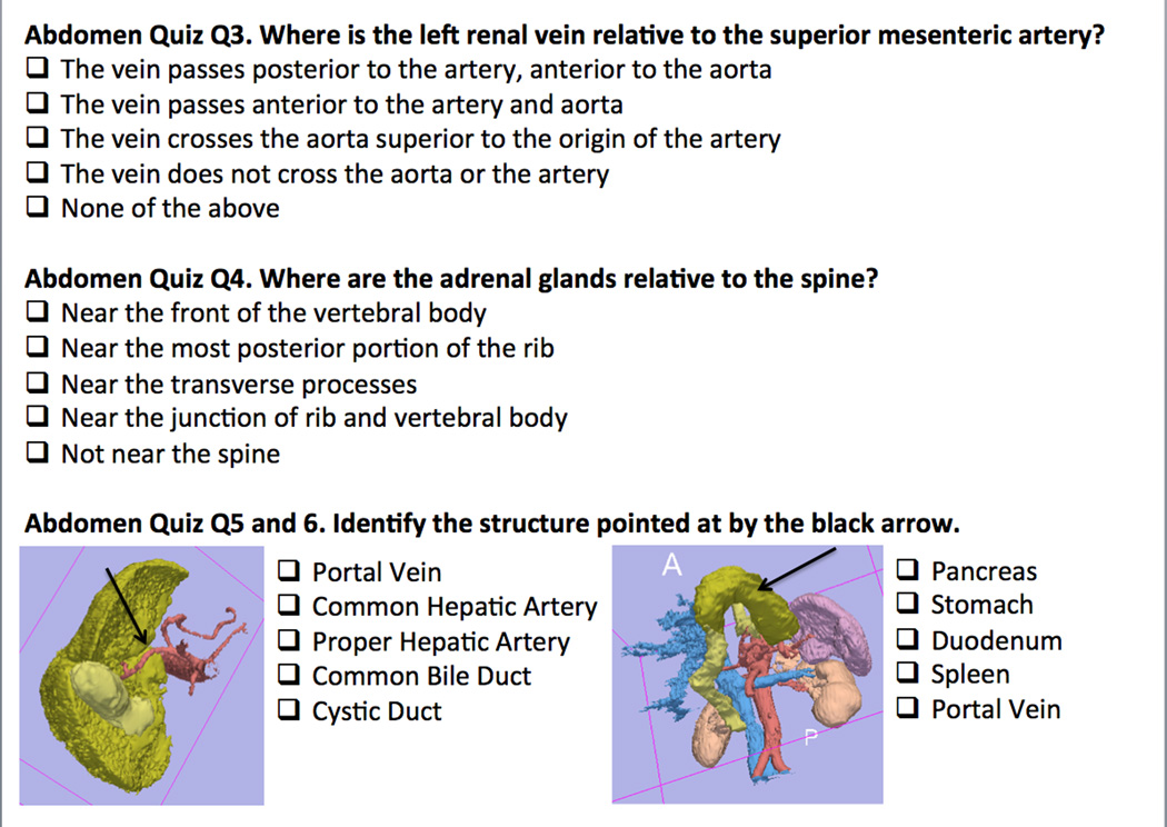
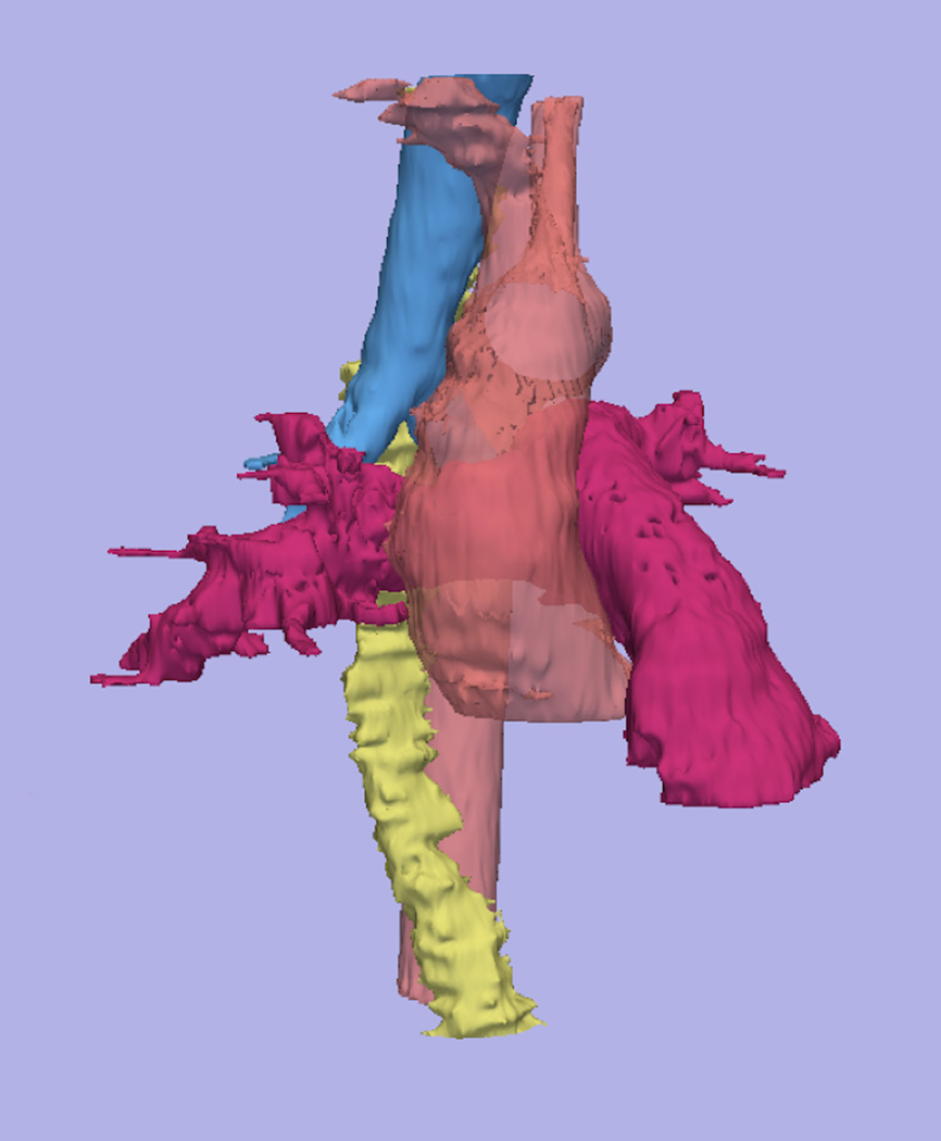
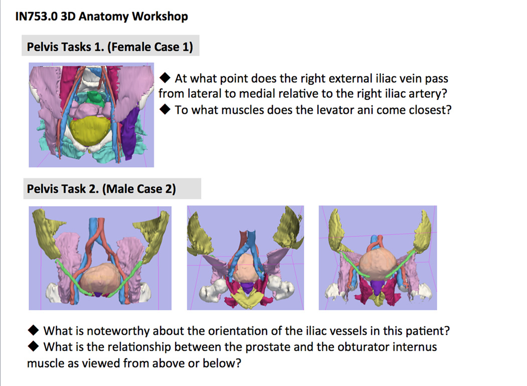
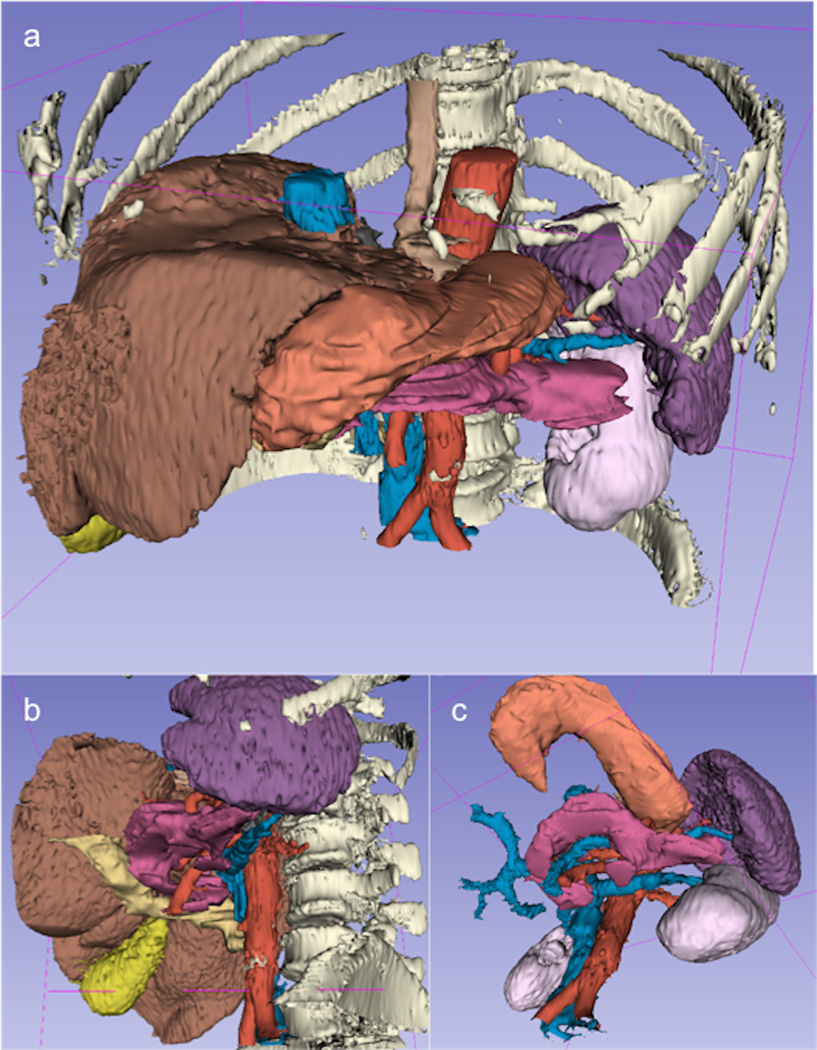
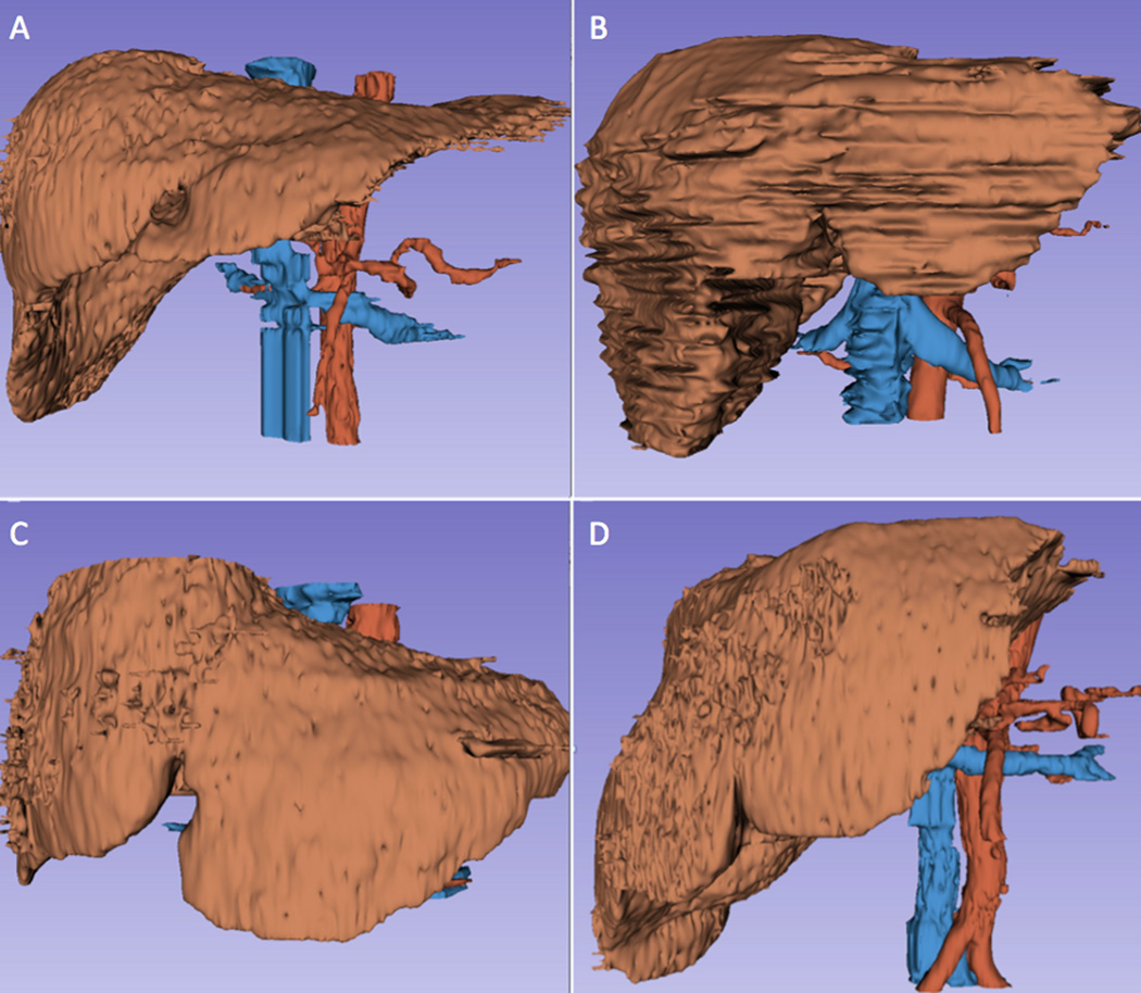
Materials and methods: A total of 196 models of anatomical structures from 16 anonymized computed tomography datasets were generated using the 3D Slicer open-source software platform. The models focused on three anatomical areas: the mediastinum, the upper abdomen, and the pelvis. Online optional quizzes were offered to first-year medical students to assess their comprehension in the areas of interest. Specific tasks were designed for students to complete using the 3D models.
Results: Scores of the quizzes confirmed a lack of understanding of 3D spatial relationships of anatomical structures despite standard instruction including dissection. Written task material and qualitative review by students suggested that interaction with 3D models led to a better understanding of the shape and spatial relationships among structures, and helped illustrate anatomical variations from one body to another.
Conclusions: The study demonstrates the feasibility of one possible approach to the generation of 3D models of the anatomy from actual patient data. The educational materials developed have the potential to supplement the teaching of complex anatomical regions and help demonstrate the anatomical variation among patients.
Keywords: 3D visualization; anatomy; open-source software.
Copyright © 2016 The Association of University Radiologists. Published by Elsevier Inc. All rights reserved.
Figures

References
-
- Vazquez R, Riesco JM, Carretero J. Reflections and Challenges in the teaching of human anatomy at the beginning of the 21st century. Eur J Anat. 2005;9(2):111–115.
-
- Duncan JS, Ayache N. Medical image analysis: progress over two decades and the challenges ahead. IEEE Trans Pattern Anal Machine Intell. 2000;22:85–105.
-
- Pommert A, Hohne KH, Plesser B, et al. Creating a high-resolution spatial/symbolic model of the inner organs based on the Visible Human. Med Image Anal. 2001;5:221–228. - PubMed
-
- Gehrmann S, Hohne KH, Linhart W, Pommert A, Tiede U, Yarar S. A detailed 3D model of the human hand. J Visualization. 2004;7:221.
Publication types
MeSH terms
Grants and funding
LinkOut - more resources
Full Text Sources
Other Literature Sources
Medical
