. 2016 Feb 22:6:21808.
doi: 10.1038/srep21808.

CD36 is a co-receptor for hepatitis C virus E1 protein attachment

Affiliations

CD36 is a co-receptor for hepatitis C virus E1 protein attachment

Jun-Jun Cheng et al. Sci Rep. .

Abstract

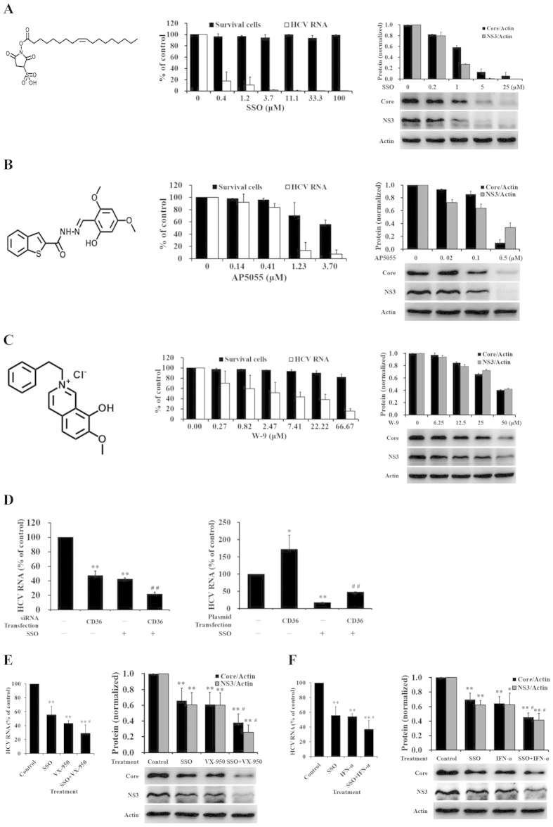
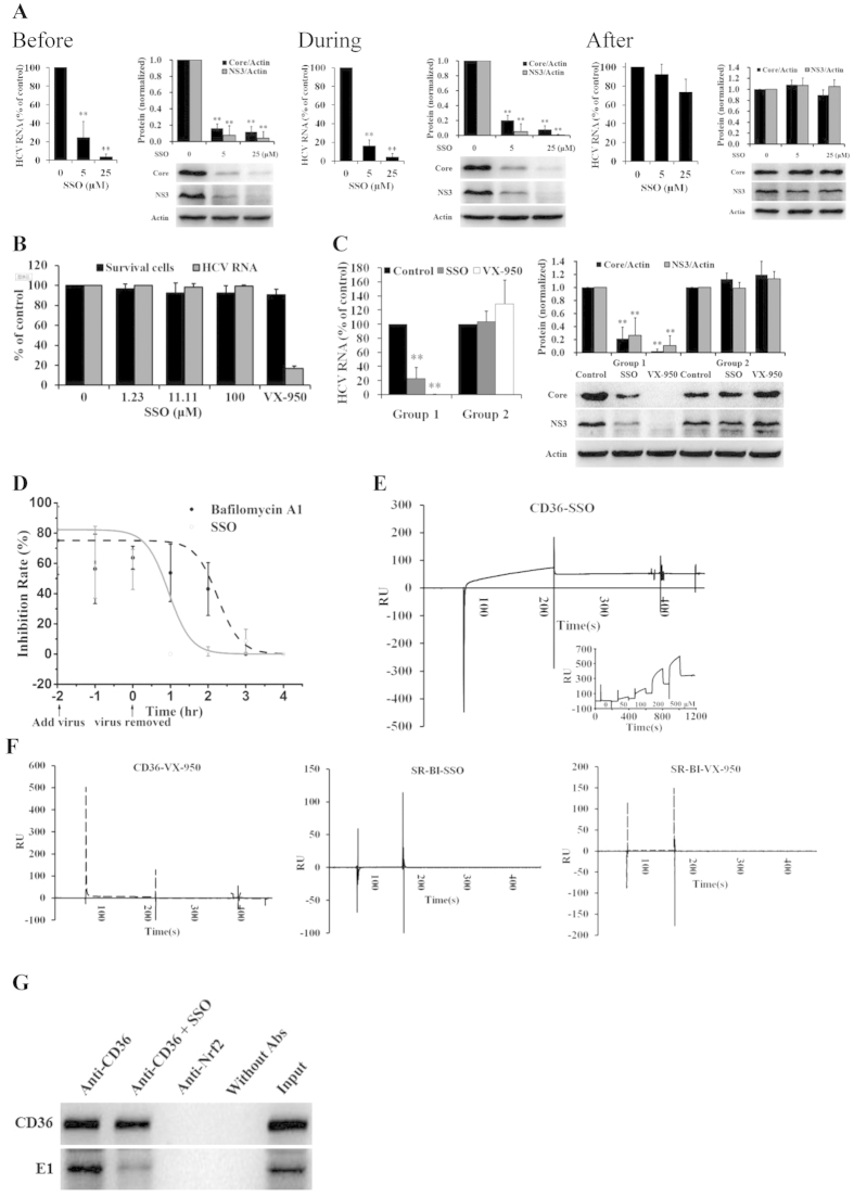
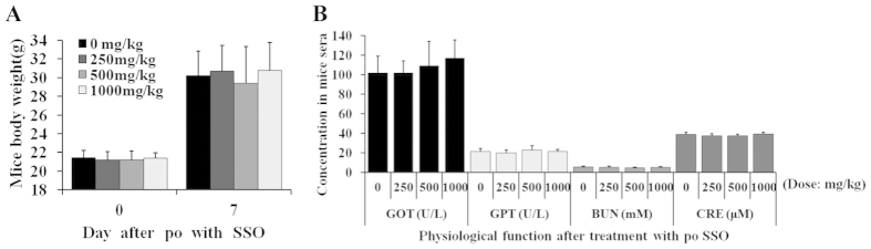
The cluster of differentiation 36 (CD36) is a membrane protein related to lipid metabolism. We show that HCV infection in vitro increased CD36 expression in either surface or soluble form. HCV attachment was facilitated through a direct interaction between CD36 and HCV E1 protein, causing enhanced entry and replication. The HCV co-receptor effect of CD36 was independent of that of SR-BI. CD36 monoclonal antibodies neutralized the effect of CD36 and reduced HCV replication. CD36 inhibitor sulfo-N-succinimidyl oleate (SSO), which directly bound CD36 but not SR-BI, significantly interrupted HCV entry, and therefore inhibited HCV replication. SSO's antiviral effect was seen only in HCV but not in other viruses. SSO in combination with known anti-HCV drugs showed additional inhibition against HCV. SSO was considerably safe in mice. Conclusively, CD36 interacts with HCV E1 and might be a co-receptor specific for HCV entry; thus, CD36 could be a potential drug target against HCV.


Figures

Figure 1
Figure 1. HCV infection increased CD36 expression in vitro.
(A) CD36 expression on naïve Huh7.5 cells was confirmed with the band at the site of exogenous CD36-HA detected with anti-HA antibody. The ratio of CD36/Actin is the average taken from 4 independent experiments (*P < 0.05 vs plasmid control (−)). (B) HCV infection increased CD36 expression on Huh7.5 cells and elevated sCD36 in culture supernatants (n = 3; *P < 0.05 and **P < 0.01 vs day 0; #P < 0.05 vs day 2). (C) CD36 expression and sCD36 secretion were increased on Huh7.5 cells infected with HCV for over 60 days (n = 3; *P < 0.05 and **P < 0.01 vs naïve control). Huh7.5 cells were infected with HCV (45IU/cell), proteins and intracellular HCV RNA were respectively detected with WB and qRT-PCR at indicated days after infection in (B,C). The protein bands presented in the figure showed the results of a representative experiment. Data presented are mean ± standard deviation. Student’s t-test was used.
Figure 2
Figure 2. HCV replication was increased by CD36.
(A) High expression of CD36 increased HCV infection and replication (n = 3). (B) Silence of CD36 decreased HCV infection and replication (n = 3). Huh7.5 cells were transfected with 0.2 μg CD36-HA plasmid (A) or with 150 pmol siRNA for CD36 (B) for 48 hrs, and then were washed and infected with HCV (150 IU/cell) for 2 hrs, followed by washing and continuously incubating. Intracellular HCV RNA and proteins were detected in 96 hrs, and the cytotoxicity of siRNAs was measured in 96-wells plate with a MTT assay in B. (C) High sCD36 in supernatants increased HCV infection (n = 3 for RNA, n = 4 for protein). (D) Low sCD36 in supernatants decreased HCV infection (n = 3 for RNA and cytotoxicity, n = 4 for protein). Huh7.5 cells were incubated with culture supernatants of Huh7.5 cells transfected with 0.2 μg CD36-HA plasmid (C) or with 150 pmol siRNA for CD36 (D) for 48 hrs, and infected with HCV (150 IU/cell) for 2 hrs, followed by washing and continuously culturing. Intracellular HCV RNA and proteins were detected in 72 hrs, and cytotoxicity was measured with a MTT assay accordingly in D. The pcDAN3.1(+) vector was used as plasmid control in A and C. The SR-BI siRNA (sc-44753) was used as positive control and siRNA-A as negative control in (B). *P < 0.05 and **P < 0.01, vs control; #P < 0.05, vs CD36 siRNA. (E) CD36 mAbs neutralized HCV infection in a dose-dependent manner (concentrations of ab17044 were 0.2, 1, and 5 μg/mL) (n = 3). (F) CD36 mAbs synergistically inhibited HCV infection with SR-BI antibody (n = 4). Huh7.5 cells were incubated with 5 μg/mL mAb for 1 hr and then infected with HCV (150 IU/cell) for 2 hrs, followed by washing and culturing. Intracellular HCV RNA was detected in 72 hrs (E,F). *P < 0.05 and **P < 0.01, vs IgG group; #,P < 0.05, vs monotherapy with ab23680 or SR-BI antibody. The mAbs code was from Abcam, Co. Ltd. (G) Cross-silencing test of CD36 and SR-BI (sc-44752), and cytotoxicity was measured with a MTT assay (n = 3 for RNA and cytotoxicity, n = 6 for protein). The protein bands presented in the figure showed the results of a representative experiment. Presented are mean ± standard deviation. Student’s t-test was used in (A–G).
Figure 3
Figure 3. CD36 directly interacted with HCV E1 protein.
(A) HCV particles bound sCD36 in culture supernatants. The culture supernatants of HCV-infected or naïve Huh7.5 cells were ultracentrifuged, and HCV viral components (Core) and sCD36 were simultaneously detected in the precipitates using western blot. (B) sCD36 bound onto HCV particles in culture supernatants (Co-IP assay). Co-IP with anti-CD36 mAb from the supernatants of HCV-infected Huh7.5 cells brought down HCV particles. (C) CD36 directly bound HCV E1 protein. Co-IP was performed with the lysates of HCV-infected Huh7.5 cells. Anti-Nrf2 antibody was used as an irrelevant negative control, and Without Abs as blank control in (B,C). (D) CD36 on cell membrane helped host to capture HCV particles (n = 3). After 48 hrs transfection with plasmid or siRNA for CD36, 293T/17 cells (resistant to HCV infection) were washed and incubated with HCV (300 IU/cell) for 3 hrs, followed by washing and then treating with lysis buffer. CD36 and HCV proteins in the lysates were analyzed with western blot. *P < 0.05, vs plasmid control (−); #P < 0.05 and ##P < 0.01, vs siRNA control (−). The protein bands presented showed the results of a representative experiment. Presented are mean ± standard deviation, and Student’s t-test was used.
Figure 4
Figure 4. CD36 inhibitors blocked HCV replication in vitro.
(AC) CD36 inhibitors prevented HCV from replicating (n = 3). Huh7.5 cells were infected with HCV (45 IU/cell) and simultaneously treated with compound SSO (A), AP5055 (B), W-9 (C) or solvent control. After 72 hrs incubation, intracellular RNA and proteins were detected, and cytotoxicity was measured with MTT assay (Middle). (D) The anti-HCV activity of SSO was affected by the level of CD36 (n = 3). Huh7.5 cells were transfected with 150 pmol siRNA for CD36 (Left) or with 0.2 μg CD36 plasmid (Right). After 48 hrs, the cells were washed and infected with HCV (150 IU/cell) and simultaneously treated with 5 μM SSO for 2 hrs, followed by washing and continuously incubating with fresh media. Intracellular HCV RNA was detected in 96 hrs. *P < 0.05 and **P < 0.01, vs siRNA (or plasmid) control plus solvent control group; ##P < 0.01, vs siRNA (or plasmid) control plus SSO group. (E) SSO in combination with VX-950 inhibited HCV replication in an additive manner (n = 3 for RNA, n = 4 for protein). (F) Increased anti-HCV effect of SSO in combination with IFN-α (n = 3 for RNA, n = 4 for protein). Huh7.5 cells were infected with HCV (45 IU/cell) and incubated with SSO (1.0 μM) or VX-950 (0.1 μM), or the combination of SSO and VX-950 (E), or incubated with IFN-α (1.0 U/mL), or the combination of SSO and IFN-α (F). Intracellular RNA and proteins were detected in 72 hrs. *P < 0.05 and **P < 0.01, vs solvent control; #P < 0.05, vs VX-950 or IFN-α alone. The protein bands showed the results of a representative experiment. Presented are mean ± standard deviation. Student’s t-test was used in (D–F).
Figure 5
Figure 5. SSO directly bound CD36 and prevented HCV from attachment on host cells.
(A) SSO only inhibited HCV replication at early stage of HCV life cycle (n = 3 for RNA, n = 4 for protein). Huh7.5 cells were treated with SSO before (at −2 hr), during (at 0 hr) or after (at 2 hr) HCV infection (150 IU/cell). After 2 hrs incubation, the supernatants were replaced with fresh culture media. Intracellular HCV RNA and proteins were detected in 72 hrs. **P < 0.01, vs solvent control. (B) SSO did not inhibit the replication of HCV sub-genome replicon in GS4.3 cells (n = 3). GS4.3 cells were treated with SSO or solvent control. After 72 hrs incubation, intracellular RNAs were detected, and cytotoxicity was measured with a MTT assay. VX-950 (1.0 μM) served as a positive control in the test. (C) SSO did not show direct inactivation on HCV (left, RNA; right, proteins) (n = 3 for RNA, n = 5 for protein). The experiment protocol is in the Method section. Intracellular HCV RNA and proteins were measured. **P < 0.01, vs solvent control in the same group. (D) Time-of-addition experiment showed that SSO prevents HCV from replication at early stage (n = 3). The experiment protocol is in the Method section. Intracellular HCV RNA was detected with qRT-PCR. Fitted lines represent sigmoidal time-dependent curves. (E) The direct interaction between CD36 and compound SSO was analyzed with BIAcore, and the interaction was in a dose-dependent manner (insert). (F) There was no direct interaction between CD36 and VX-950 (left), or SSO and SR-BI (middle), or VX-950 and SR-BI (right), in the BIAcore analysis. The protein bands presented showed the results of a representative experiment. Presented is mean ± standard deviation, and Student’s t-test was used in (A–D). (G) The interaction between CD36 and HCV E1 protein was disrupted by SSO. Co-IP assay was performed with the lysates of HCV-infected Huh7.5 cells and 20 μM SSO was added additionally. Anti-Nrf2 antibody was used as an irrelevant negative control, and Without Abs as blank control.
Figure 6
Figure 6. Safety of SSO in mice.
Oral administration of SSO (single dosing) in mice (n = 10; ♂ × 5 and ♀ × 5) caused no change in survival and body weight (A); liver and kidney function was without change as well (B). GOT, glutamate-oxaloacetate transaminase (U/L); GPT, glutamate-pyruvate transaminase (U/L); BUN, blood urea nitrogen (mM); CRE, creatine (μM).

Similar articles

Cited by

References

    1. Dubuisson J. & Cosset F. L. Virology and cell biology of the hepatitis C virus life cycle: an update. J. Hepatol. 61, S3–S13 (2014). - PubMed
    1. Lindenbach B. D. & Rice C. M. The ins and outs of hepatitis C virus entry and assembly. Nat. Rev. Microbiol. 11, 688–700 (2013). - PMC - PubMed
    1. Lin L. T. et al. Broad-spectrum antiviral activity of chebulagic acid and punicalagin against viruses that use glycosaminoglycans for entry. BMC Microbiol. 13, 187 (2013). - PMC - PubMed
    1. Albecka A. et al. Role of low-density lipoprotein receptor in the hepatitis C virus life cycle. Hepatology 55, 998–1007 (2012). - PubMed
    1. Zona L. et al. CD81-receptor associations–impact for hepatitis C virus entry and antiviral therapies. Viruses 6, 875–892 (2014). - PMC - PubMed

Publication types

MeSH terms