. 2016 Mar 7;26(5):678-85.
doi: 10.1016/j.cub.2015.12.073. Epub 2016 Feb 18.

Chromosome Cohesion Established by Rec8-Cohesin in Fetal Oocytes Is Maintained without Detectable Turnover in Oocytes Arrested for Months in Mice

Affiliations

Chromosome Cohesion Established by Rec8-Cohesin in Fetal Oocytes Is Maintained without Detectable Turnover in Oocytes Arrested for Months in Mice

Sabrina Burkhardt et al. Curr Biol. .

Abstract

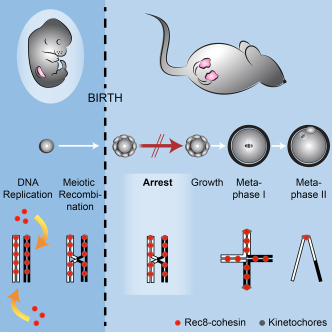
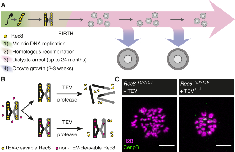
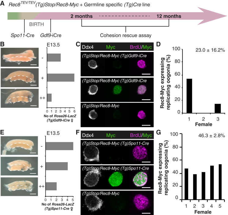
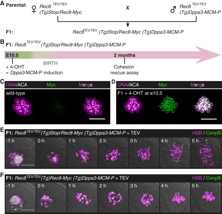
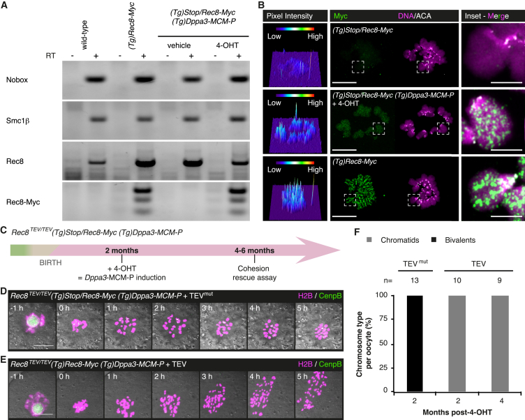
Sister chromatid cohesion mediated by the cohesin complex is essential for chromosome segregation in mitosis and meiosis [1]. Rec8-containing cohesin, bound to Smc3/Smc1α or Smc3/Smc1β, maintains bivalent cohesion in mammalian meiosis [2-6]. In females, meiotic DNA replication and recombination occur in fetal oocytes. After birth, oocytes arrest at the prolonged dictyate stage until recruited to grow into mature oocytes that divide at ovulation. How cohesion is maintained in arrested oocytes remains a pivotal question relevant to maternal age-related aneuploidy. Hypothetically, cohesin turnover regenerates cohesion in oocytes. Evidence for post-replicative cohesion establishment mechanism exists, in yeast and invertebrates [7, 8]. In mouse fetal oocytes, cohesin loading factor Nipbl/Scc2 localizes to chromosome axes during recombination [9, 10]. Alternatively, cohesion is maintained without turnover. Consistent with this, cohesion maintenance does not require Smc1β transcription, but unlike Rec8, Smc1β is not required for establishing bivalent cohesion [11, 12]. Rec8 maintains cohesion without turnover during weeks of oocyte growth [3]. Whether the same applies to months or decades of arrest is unknown. Here, we test whether Rec8 activated in arrested mouse oocytes builds cohesion revealed by TEV cleavage and live-cell imaging. Rec8 establishes cohesion when activated during DNA replication in fetal oocytes using tamoxifen-inducible Cre. In contrast, no new cohesion is detected when Rec8 is activated in arrested oocytes by tamoxifen despite cohesin synthesis. We conclude that cohesion established in fetal oocytes is maintained for months without detectable turnover in dictyate-arrested oocytes. This implies that women's fertility depends on the longevity of cohesin proteins that established cohesion in utero.

Keywords: cohesin; meiosis; oocytes.


Figures

None
Graphical abstract
Figure 1
Figure 1
Probing Cohesion Maintenance during Mammalian Female Meiosis Using a Functional Cohesion Assay (A) Mammalian female meiosis can be structured into four stages: (1) meiotic DNA replication in which sister chromatid cohesion is presumably established, (2) meiotic recombination in which reciprocal recombination of homologous chromosomes (black and gray) results in crossovers that manifest as chiasmata, (3) the prolonged resting state at the dictyate stage of prophase I after birth, and (4) the growing phase that starts when an oocyte is recruited from the resting pool and leads to formation of a mature oocyte. The duration of the dictyate arrest and growing phase correspond to mouse. Schematic is not drawn to scale. (B) Schematic of cohesion rescue assay. TEV-cleavable Rec8 (yellow) establishes and maintains bivalent cohesion. Non-TEV-cleavable Rec8 (red) is activated after meiotic cohesion establishment. Cleavage by TEV protease reveals whether de novo expressed non-TEV-cleavable Rec8 is able to build functional cohesive structures: cleavage of bivalents into single chromatids indicates no functional loading, while resistance to TEV cleavage reveals functional cohesive structures entrapping sister DNA molecules. (C) Rec8TEV/TEV oocytes are microinjected with mRNA encoding H2B-mCherry, CenpB-EGFP, and TEV protease. Confocal time-lapse microscopy allows scoring of chromosome type at metaphase I (5 hr post-GVBD). TEV protease efficiently converts bivalents to chromatids, which are detected as at least 72 single chromatids and no bivalents (oocytes analyzed n = 40), while no cleavage of all 20 bivalents is observed using mutant TEV protease (TEVmut; oocytes analyzed n = 16). Scale bar, 10 μm. See also Figure S1.
Figure 2
Figure 2
Gdf9-iCre and Spo11-Cre Activate Rec8-Myc during Meiotic DNA Replication (A) Interpretation of the cohesion rescue assay requires activation of the Rec8-Myc transgene after meiotic DNA replication (green). Thus, Gdf9-iCre or Spo11-Cre was used to activate Rec8-Myc in oocytes shortly after birth (dictyate stage, pink) or during homologous recombination (beige), respectively. (B) Timely deletion analysis of Gdf9-iCre. Scoring of X-gal positive cells in Rosa26-LacZ (Tg)Gdf9-iCre embryonic ovaries at E13.5 according to classification into negative (−), weakly positive (+), and positive (++); n = 10 female embryos. The dashed line indicates the gonad. Scale bar, 1 mm. (C and D) Gdf9-iCre activates Rec8-Myc during meiotic S phase. Embryonic day E13.5 (Tg)Stop/Rec8-Myc (Tg)Gdf9-iCre embryonic ovaries were scored for Rec8-Myc expressing replicating (BrdU-positive) oogonia identified by germ cell-specific cytoplasmic Ddx4 staining; n = 3 females. No Myc signal was observed in oogonia from control (Tg)Stop/Rec8-Myc females; n = 2 females. (C) Representative images; scale bar, 10 μm. (D) Quantification for (Tg)Stop/Rec8-Myc (Tg)Gdf9-iCre embryonic ovaries; n > 100 cells per female. Mean ± SEM given. (E–G) Timely deletion analysis of Spo11-Cre. (E) Scoring of X-gal positive cells in Rosa26-LacZ (Tg)Spo11-Cre embryonic ovaries at E13.5 according classification into negative (–), weakly positive (+), and positive (++); n = 11 female embryos. The dashed line indicates the gonad. Scale bar, 1 mm. (F and G) Spo11-Cre activates Rec8-Myc during meiotic S phase. Embryonic day E14.5 (Tg)Stop/Rec8-Myc (Tg)Spo11-Cre embryonic ovaries were scored for Rec8-Myc expressing replicating (BrdU-positive) oogonia identified by germ cell-specific cytoplasmic Ddx4 staining; n = 5 females. No Myc signal was observed in oogonia from control (Tg)Stop/Rec8-Myc females; n = 3 females. (F) Representative images; scale bar, 10 μm. (G) Quantification for (Tg)Stop/Rec8-Myc (Tg)Spo11-Cre embryonic ovaries; n > 500 cells per female. Mean ± SEM given. See also Figure S2 and Table S1.
Figure 3
Figure 3
Rec8 Activated in Fetal Oocytes Builds Cohesive Structures (A) Mating scheme to obtain female F1 offspring of the required genotype Rec8TEV/TEV(Tg)Stop/Rec8-Myc (Tg)Dppa3-MCM-P. (B) Activation of Rec8-Myc during meiotic S phase in fetal oocytes. Pregnant Rec8TEV/TEV(Tg)Stop/Rec8-Myc females are injected with 4-OHT on embryonic day E10.5 to induce deletion by Dppa3-MCM-P in embryos. Oocytes are isolated from F1 females with the appropriate genotype. Green, meiotic DNA replication; beige, homologous recombination; pink, dictyate stage. (C and D) Metaphase I chromosome spread showing localization of Rec8-Myc to bivalent chromosomes in oocytes from (C) wild-type and (D) F1 Rec8TEV/TEV(Tg)Stop/Rec8-Myc (Tg)Dppa3-MCM-P female; oocytes analyzed n = 9 and n = 7, respectively. Centromeres are marked by anti-centromere antibody (ACA). Scale bar, 20 μm. (E and F) Representative still images of oocytes isolated from F1 Rec8TEV/TEV(Tg)Stop/Rec8-Myc (Tg)Dppa3-MCM-P females microinjected with mRNA encoding H2B-mCherry, CenpB-EGFP, and TEV protease. (E) Bivalents are converted to chromatids in cells without Stop cassette deletion; n = 17 oocytes. (F) Bivalents are retained in oocytes with successful deletion of the Stop cassette; n = 5 oocytes. Genotype of single cells was confirmed after live-cell imaging. Oocytes were obtained from >3 females. Scale bar, 20 μm. See also Figure S3 and Table S1.
Figure 4
Figure 4
Bivalent Cohesion Is Maintained without Detectable Turnover of Rec8-Cohesive Structures for Months during the Dictyate Arrest (A) Rec8-Myc is transcribed in adult ovary. Detection of mRNA for Nobox, Smc1β, Rec8, and Rec8-Myc by RT-PCR from adult ovary. RT, reverse transcriptase. See also Figure S4. (B) Rec8-Myc is synthesized in oocytes from adult Rec8TEV/TEV(Tg)Stop/Rec8-Myc (Tg)Dppa3-MCM-P female analyzed 2 months post-4-OHT. Metaphase I chromosome spread showing expression and localization of Rec8-Myc to bivalent chromosomes. Left panel shows 3D surface plots of Rec8-Myc pixel fluorescence intensities. Centromeres are marked by ACA. Oocytes analyzed from top to bottom: n = 11, 7, 16. Inset has been brightness and contrast enhanced equally in all three panels. Scale bar, 20 μm; inset scale bar, 5 μm. (C) Timing of cohesion rescue assay utilizing Dppa3-MCM-P to activate Rec8-Myc in adult female mice. Green, meiotic DNA replication; beige, homologous recombination; pink, dictyate stage. (D and E) Representative still images of oocytes isolated from Rec8TEV/TEV(Tg)Stop/Rec8-Myc (Tg)Dppa3-MCM-P females microinjected with mRNA encoding H2B-mCherry, CenpB-EGFP, and TEV protease. (D) Bivalents are retained in oocytes microinjected with mutant TEV mRNA. (E) TEV protease converts bivalents to chromatids in cells with successful Stop cassette deletion. Single-cell genotyping was performed after live-cell imaging. Scale bar, 20 μm. (F) Quantification of cohesion rescue assay in oocytes from Rec8TEV/TEV(Tg)Stop/Rec8-Myc (Tg)Dppa3-MCM-P females with successful Stop cassette deletion used 2 or 4 months post-4-OHT treatment. Oocytes were obtained from >3 females; n = number of oocytes analyzed. See also Figure S3; Table S1; Movies S1 and S2.

Comment in

References

    1. Nasmyth K., Haering C.H. Cohesin: its roles and mechanisms. Annu. Rev. Genet. 2009;43:525–558. - PubMed
    1. Watanabe Y., Nurse P. Cohesin Rec8 is required for reductional chromosome segregation at meiosis. Nature. 1999;400:461–464. - PubMed
    1. Tachibana-Konwalski K., Godwin J., van der Weyden L., Champion L., Kudo N.R., Adams D.J., Nasmyth K. Rec8-containing cohesin maintains bivalents without turnover during the growing phase of mouse oocytes. Genes Dev. 2010;24:2505–2516. - PMC - PubMed
    1. Lee J., Iwai T., Yokota T., Yamashita M. Temporally and spatially selective loss of Rec8 protein from meiotic chromosomes during mammalian meiosis. J. Cell Sci. 2003;116:2781–2790. - PubMed
    1. Revenkova E., Eijpe M., Heyting C., Gross B., Jessberger R. Novel meiosis-specific isoform of mammalian SMC1. Mol. Cell. Biol. 2001;21:6984–6998. - PMC - PubMed

Publication types