

The microRNA-23b/-27b cluster suppresses prostate cancer metastasis via Huntingtin-interacting protein 1-related
- PMID: 26898757
- PMCID: PMC5770234
- DOI: 10.1038/onc.2016.6
The microRNA-23b/-27b cluster suppresses prostate cancer metastasis via Huntingtin-interacting protein 1-related
Abstract
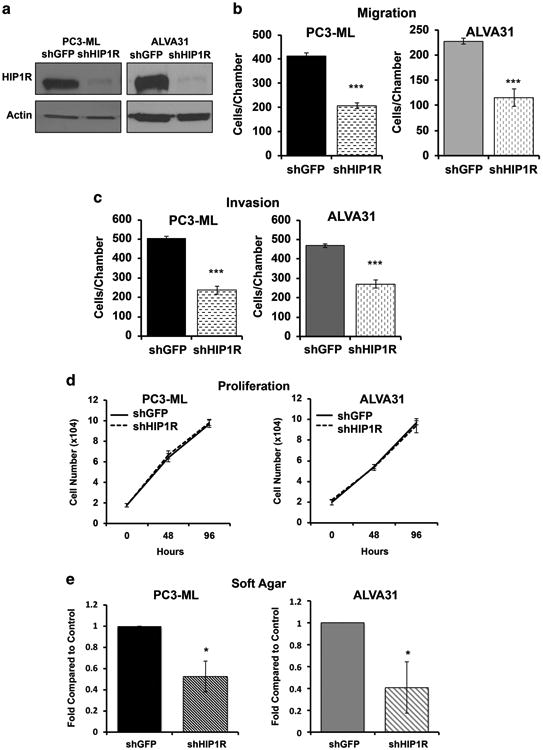
Deregulation of microRNAs (miRs) contributes to progression and metastasis of prostate and other cancers. miR-23b and -27b, encoded in the same miR cluster (miR-23b/-27b), are downregulated in human metastatic prostate cancer compared with primary tumors and benign tissue. Expression of miR-23b/-27b decreases prostate cancer cell migration, invasion and results in anoikis resistance. Conversely, antagomiR-mediated miR-23b and -27b silencing produces the opposite result in a more indolent prostate cancer cell line. However, neither miR-23b/-27b expression or inhibition impacts prostate cancer cell proliferation suggesting that miR-23b/-27b selectively suppresses metastasis. To examine the effects of miR-23b/-27b on prostate cancer metastasis in vivo, orthotopic prostate xenografts were established using aggressive prostate cancer cells transduced with miR-23b/-27b or non-targeting control miRNA. Although primary tumor formation was similar between miR-23b/-27b-transduced cells and controls, miR-23b/-27b expression in prostate cancer cells decreased seminal vesicle invasion and distant metastases. Gene-expression profiling identified the endocytic adaptor, Huntingtin-interacting protein 1-related (HIP1R) as being downregulated by miR-23b/-27b. Increased HIP1R expression in prostate cancer cells inversely phenocopied the effects of miR-23b/-27b overexpression on migration, invasion and anchorage-independent growth. HIP1R rescued miR-23b/-27b-mediated repression of migration in prostate cancer cells. HIP1R mRNA levels were decreased in seminal vesicle tissue from mice bearing miR-23b/-27b-transduced prostate cancer cell xenografts compared with scrambled controls, suggesting HIP1R is a key functional target of miR-23b/-27b. In addition, depletion of HIP1R led to a more rounded, less mesenchymal-like cell morphology, consistent with decreased metastatic properties. Together, these data demonstrate that the miR-23b/-27b cluster functions as a metastasis-suppressor by decreasing HIP1R levels in pre-clinical models of prostate cancer.
Conflict of interest statement
Figures


Similar articles
-
The microRNA -23b/-27b cluster suppresses the metastatic phenotype of castration-resistant prostate cancer cells.PLoS One. 2012;7(12):e52106. doi: 10.1371/journal.pone.0052106. Epub 2012 Dec 26. PLoS One. 2012. PMID: 23300597 Free PMC article.
-
Dual regulation of receptor tyrosine kinase genes EGFR and c-Met by the tumor-suppressive microRNA-23b/27b cluster in bladder cancer.Int J Oncol. 2015 Feb;46(2):487-96. doi: 10.3892/ijo.2014.2752. Epub 2014 Nov 14. Int J Oncol. 2015. PMID: 25405368 Free PMC article.
-
MicroRNA-23b and microRNA-27b plus flutamide treatment enhances apoptosis rate and decreases CCNG1 expression in a castration-resistant prostate cancer cell line.Tumour Biol. 2018 Nov;40(11):1010428318803011. doi: 10.1177/1010428318803011. Tumour Biol. 2018. PMID: 30400755
-
Promising therapeutic role of miR-27b in tumor.Tumour Biol. 2017 Mar;39(3):1010428317691657. doi: 10.1177/1010428317691657. Tumour Biol. 2017. PMID: 28351320 Review.
-
Functional significance of aberrantly expressed microRNAs in prostate cancer.Int J Urol. 2015 Mar;22(3):242-52. doi: 10.1111/iju.12700. Epub 2015 Jan 20. Int J Urol. 2015. PMID: 25599923 Review.
Cited by
-
Flurbiprofen inhibits cell proliferation in thyroid cancer through interrupting HIP1R-induced endocytosis of PTEN.Eur J Med Res. 2022 Feb 24;27(1):29. doi: 10.1186/s40001-022-00658-3. Eur J Med Res. 2022. PMID: 35209947 Free PMC article.
-
Knockdown of microRNA-214-3p Promotes Tumor Growth and Epithelial-Mesenchymal Transition in Prostate Cancer.Cancers (Basel). 2021 Nov 23;13(23):5875. doi: 10.3390/cancers13235875. Cancers (Basel). 2021. PMID: 34884984 Free PMC article.
-
HIP1R acts as a tumor suppressor in gastric cancer by promoting cancer cell apoptosis and inhibiting migration and invasion through modulating Akt.J Clin Lab Anal. 2020 Sep;34(9):e23425. doi: 10.1002/jcla.23425. Epub 2020 Jun 16. J Clin Lab Anal. 2020. PMID: 32548851 Free PMC article.
-
Bioinformatics-Assisted Extraction of All PCa miRNAs and their Target Genes.Microrna. 2024;13(1):33-55. doi: 10.2174/0122115366253242231020053221. Microrna. 2024. PMID: 38284737
-
The Functional Role of Prostate Cancer Metastasis-related Micro-RNAs.Cancer Genomics Proteomics. 2019 Jan-Feb;16(1):1-19. doi: 10.21873/cgp.20108. Cancer Genomics Proteomics. 2019. PMID: 30587496 Free PMC article. Review.
References
-
- Jemal A, Siegel R, Xu J, Ward E. Cancer statistics, 2010. CA Cancer J Clin. 2010;60:277–300. - PubMed
-
- Lagos-Quintana M, Rauhurt R, Lendeckel W, Tuschi T. Identification of novel genes coding for small expressed RNAs. Science. 2001;294:853–858. - PubMed
-
- Dalmay T, Edwards DR. MicroRNAs and the hallmarks of cancer. Oncogene. 2006;25:6170–6175. - PubMed
MeSH terms
Substances
Grants and funding
LinkOut - more resources
Full Text Sources
Other Literature Sources
Medical
Molecular Biology Databases
Research Materials
