. 2016 Apr;131(4):525-37.
doi: 10.1007/s00401-016-1546-0. Epub 2016 Feb 22.

Phosphorylation of the amyloid β-peptide at Ser26 stabilizes oligomeric assembly and increases neurotoxicity

Affiliations

Phosphorylation of the amyloid β-peptide at Ser26 stabilizes oligomeric assembly and increases neurotoxicity

Sathish Kumar et al. Acta Neuropathol. 2016 Apr.

Abstract

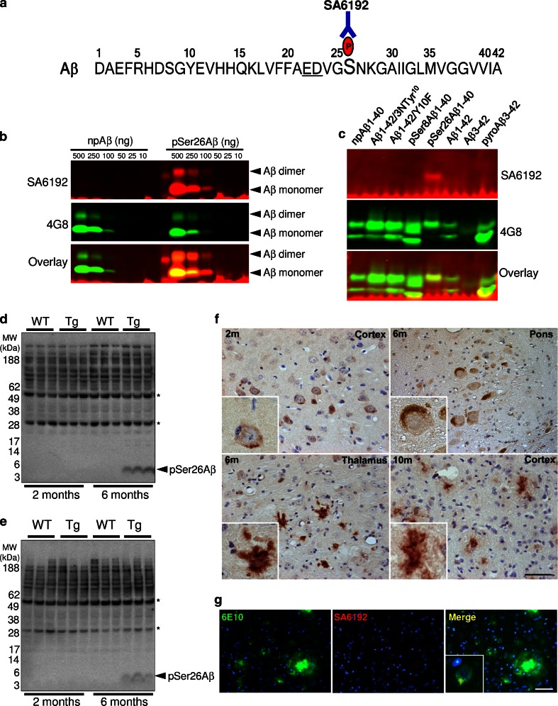
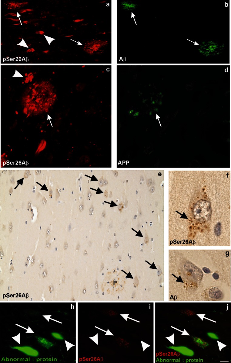
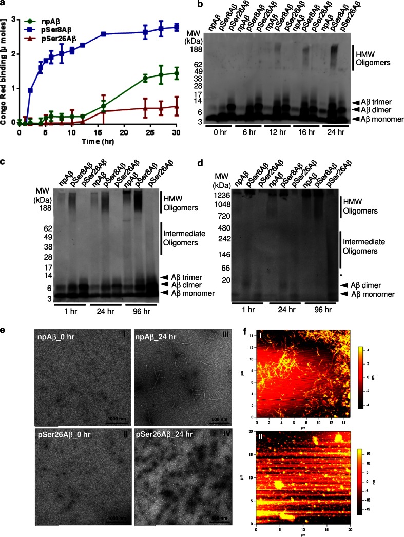
Aggregation and toxicity of the amyloid β-peptide (Aβ) are considered as critical events in the initiation and progression of Alzheimer's disease (AD). Recent evidence indicated that soluble oligomeric Aβ assemblies exert pronounced toxicity, rather than larger fibrillar aggregates that deposit in the forms of extracellular plaques. While some rare mutations in the Aβ sequence that cause early-onset AD promote the oligomerization, molecular mechanisms that induce the formation or stabilization of oligomers of the wild-type Aβ remain unclear. Here, we identified an Aβ variant phosphorylated at Ser26 residue (pSer26Aβ) in transgenic mouse models of AD and in human brain that shows contrasting spatio-temporal distribution as compared to non-phosphorylated Aβ (npAβ) or other modified Aβ species. pSer26Aβ is particularly abundant in intraneuronal deposits at very early stages of AD, but much less in extracellular plaques. pSer26Aβ assembles into a specific oligomeric form that does not proceed further into larger fibrillar aggregates, and accumulates in characteristic intracellular compartments of granulovacuolar degeneration together with TDP-43 and phosphorylated tau. Importantly, pSer26Aβ oligomers exert increased toxicity in human neurons as compared to other known Aβ species. Thus, pSer26Aβ could represent a critical species in the neurodegeneration during AD pathogenesis.

Keywords: Alzheimer’s disease; Amyloid oligomer; Granulovacuolar degeneration; Intraneuronal Abeta; Phosphorylation; Protein aggregation.


Figures

Fig. 1
Fig. 1
Specific detection of Aβ phosphorylated at Ser26 in transgenic mouse models of AD. a Amino acid sequence of human Aβ indicating the phosphorylation site at Ser26. Underlined glutamic acid (E) and aspartic acid (D) residues comprise a consensus phosphorylation sequence for casein kinase 1. b, c SA6192 antibody specifically detects pSer26Aβ in immunoblotting without cross-reactivity against other post-translationally modified or non-modified Aβ variants (c). Generic 4G8 antibody recognizes non-modified and all the modified Aβ variants (b, c). d, e WB analysis of sucrose (d) and SDS (e) fractions of 2- and 6-months-old APP/PS1KI (Tg) and non-Tg (WT) mouse brain homogenates revealed the presence of pSer26Aβ in vivo. SA6192 showed no reactivity with endogenous mouse APP in non-transgenic mice, further demonstrating the specificity of this antibody (Supplementary Fig. 1a, b). The bands indicated by asterisks likely represent heavy and light chains of endogenous immunoglobulins. f Immunohistochemical staining of 2-, 6-, and 10-month-old APP/PS1KI mouse brain tissues with SA6192 antibody demonstrates the occurrence of intraneuronal (2 and 6 months) and extracellular (10 months) pSer26Aβ deposits in different brain regions. g Double-labelling with 6E10 (green) and SA6192 (red) revealed intraneuronal immunoreactivity of SA6192. The inset in the merged image shows a higher magnification of 6E10 and SA6192 co-localization (yellow) within the cell body of a cortical neuron. Scale bars f and g 50 μm
Fig. 2
Fig. 2
Detection of intraneuronal pSer26Aβ aggregates and GVDs in human AD brains. Detection of pSer26Aβ in extracellular (arrows) and intraneuronal (arrowheads) deposits in the hippocampal CA1 subfield (a, c, e, f, g, i). The extracellular Aβ plaques are co-stained with anti-Aβ17–24 (4G8) (b), and anti-APP antibody (22C11) (d).The central amyloid core is stained with SA6192 and 4G8 but not with anti-APP indicating the co-deposition of pSer26Aβ together with non-phosphorylated Aβ in plaques (Supplementary Fig. 5a–c). Note the intraneuronal globular aggregates reactivity is selectively observed with pSer26Aβ (arrowhead in a, c), but not with APP antibodies. Immunohistochemical analysis demonstrates strong intraneuronal granular cytoplasmic pSer26Aβ inclusions (arrows in e), and only weakly stained extracellular pSer26Aβ-positive plaques (P in e) (Supplementary Fig. 4d). These granular inclusions exhibit the morphological pattern of granulovacuolar degeneration (GVD) and most frequently occur in the CA1-subiculum area of the hippocampal formation (arrow in f). GVD was also detected by anti-Aβ17–24 staining (arrow in g). pSer26Aβ-positive GVD lesions colocalized with abnormal-phosphorylated τ in neurons (arrows in hj). Note that neurofibrillary tangles were not labelled with anti-pSer26Aβ antibody (arrowhead in hj). The panels in this figure are representative images from 4 different AD brains (a, b case # 7; c, d case # 3, eg case # 1 and h, i case # 5 of supplementary Table 2). Scale bars a and b 50 µm; c and d 30 µm; e 20 µm; f and g 5 µm; hj 20 µm
Fig. 3
Fig. 3
pSer26Aβ selectively forms oligomers without fibril formation. a Congo Red (CR) binding assay showing the decreased CR dye binding to pSer26Aβ as compared to npAβ and pSer8Aβ peptides. b SDS-PAGE and Western immunoblot detection of Aβ variants after different times of aggregation (see also supplementary Fig. 7). SDS-PAGE (c) and native-PAGE (d) analysis of the aggregates collected at different incubation time demonstrates the lack of HMW pSer26Aβ assemblies, even after prolonged incubation time (96 h). Monoclonal 82E1 antibody was used for immunoblotting. e Transmission electron microscopy (TEM) images demonstrate granular non-aggregated structures of npAβ and pSer26Aβ peptide samples at 0 h (e I, II). After 24 h of incubation, mature fibrils are only seen with npAβ (e III), whereas pSer26Aβ predominantly shows spherical non-fibrillar chain-like globular structures (e IV). f Atomic force microscopy (AFM) images of npAβ and pSer26Aβ after 24 h of aggregation further confirm the formation of fibrillar aggregates of npAβ (f, i) and non-fibrillar globular assemblies of pSer26Aβ peptide (f II, see also Supplementary Fig. 8)
Fig. 4
Fig. 4
Increased cytotoxicity of pSer26Aβ. Human neuroblastoma cells (a) and embryonic stem cell-derived neurons (hESNs) (b) were incubated with npAβ (Aβ1–42) and pseudophosphorylated (AβS26D) peptides. Cell viability was analysed by the Presto Blue assay. Both Aβ variants (Aβ1–42 and Aβ26D) did not exert toxicity at lower concentration (1 µM) in neuroblastoma cells. At concentrations of 10 µM, the pseudophosphorylated AβS26D variant was more toxic than the unmodified peptide. (a). In hESNs, AβS26D was already toxic at a 1 µM concentration, where unmodified Aβ had no effect. At 10 µM, AβS26D also exterted pronounced toxicity as compared to unmodified Aβ (b). (p < 0.01; red stars indicate statistical significance of the indicated bar graphs versus vehicle controls; black stars indicate statistical significance between the indicated bar graph pairs; mean ± standard error of the mean (SEM), n = 18 replicates from 3 independent experiments). STS staurosporine treatment (positive control), BTC buffer treatment control (similar volume of PBS without Aβ), NTC non-treated control (no addition of PBS to culture media), N.S. not significant. c, d Dot blotting of npAβ, pSer8Aβ and pSer26Aβ variants collected at the indicated time periods of incubation (0, 2, 6, 12 and 24 h) with conformation-dependent anti-amyloid oligomer-specific A11 (c upper panel) and OC (d upper panel) antibodies. Reprobing of the membranes with generic 82E1 antibody confirms the presence of Aβ variants (c, d lower panels). e Native-PAGE of the npAβ, pSer8Aβ and pSer26Aβ samples shows the kinetic differences in formation intermediate and HMW Aβ assemblies. Mouse monoclonal 82E1 antibody was used for probing the blots. Asterisk indicates trimeric/tetrameric Aβ assemblies. f Aggregates of npAβ, pSer8Aβ and pSer26Aβ were added to induced pluripotent stem cell (iPSC)-derived neurons and incubated for 50 h.The most cytotoxic species observed were the pSer26Aβ aggregates after 24 h of aggregation (p < 0.01; red stars signify statistical significance of the indicated bar graphs versus buffer controls; black stars signify statistical significance between the indicated bar graph pairs; mean ± SEM)

Similar articles

Cited by

References

    1. Baglioni S, Casamenti F, Bucciantini M, Luheshi LM, Taddei N, Chiti F, Dobson CM, Stefani M. Prefibrillar amyloid aggregates could be generic toxins in higher organisms. J Neurosci. 2006;26:8160–8167. doi: 10.1523/JNEUROSCI.4809-05.2006. - DOI - PMC - PubMed
    1. Baumketner A, Bernstein SL, Wyttenbach T, Lazo ND, Teplow DB, Bowers MT, Shea JE. Structure of the 21–30 fragment of amyloid beta-protein. Protein Sci. 2006;15:1239–1247. doi: 10.1110/ps.062076806. - DOI - PMC - PubMed
    1. Bemporad F, Chiti F. Protein misfolded oligomers: experimental approaches, mechanism of formation, and structure–toxicity relationships. Chem Biol. 2012;19:315–327. doi: 10.1016/j.chembiol.2012.02.003. - DOI - PubMed
    1. Benilova I, Karran E, De SB. The toxic Abeta oligomer and Alzheimer’s disease: an emperor in need of clothes. Nat Neurosci. 2012;15:349–357. doi: 10.1038/nn.3028. - DOI - PubMed
    1. Billings LM, Oddo S, Green KN, McGaugh JL, LaFerla FM. Intraneuronal Abeta causes the onset of early Alzheimer’s disease-related cognitive deficits in transgenic mice. Neuron. 2005;45:675–688. doi: 10.1016/j.neuron.2005.01.040. - DOI - PubMed

Publication types

MeSH terms

LinkOut - more resources