

Sclerosing bone dysplasias: genetic, clinical and radiology update of hereditary and non-hereditary disorders
- PMID: 26898950
- PMCID: PMC5258139
- DOI: 10.1259/bjr.20150349
Sclerosing bone dysplasias: genetic, clinical and radiology update of hereditary and non-hereditary disorders
Abstract
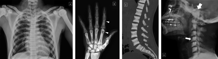
There is a wide variety of hereditary and non-hereditary bone dysplasias, many with unique radiographic findings. Hereditary bony dysplasias include osteopoikilosis, osteopathia striata, osteopetrosis, progressive diaphyseal dysplasia, hereditary multiple diaphyseal sclerosis and pyknodysostosis. Non-hereditary dysplasias include melorheostosis, intramedullary osteosclerosis and overlap syndromes. Although many of these dysplasias are uncommon, radiologists should be familiar with their genetic, clinical and imaging findings to allow for differentiation from acquired causes of bony sclerosis. We present an overview of hereditary and non-hereditary bony dysplasias with focus on the pathogenesis, clinical and radiographic findings of each disorder.
Figures


Similar articles
-
Sclerosing bone dysplasias: review and differentiation from other causes of osteosclerosis.Radiographics. 2011 Nov-Dec;31(7):1865-82. doi: 10.1148/rg.317115093. Radiographics. 2011. PMID: 22084176 Review.
-
Sclerosing bone dysplasias: genetic and radioclinical features.Eur Radiol. 2000;10(9):1423-33. doi: 10.1007/s003300000495. Eur Radiol. 2000. PMID: 10997431 Review.
-
Sclerosing bone dysplasias: a pictorial essay.Radiol Bras. 2025 Jan 6;57:e20240058. doi: 10.1590/0100-3984.2024.0058-en. eCollection 2024 Jan-Dec. Radiol Bras. 2025. PMID: 39822959 Free PMC article.
-
Sclerosing bone dysplasias--a target-site approach.Skeletal Radiol. 1991;20(8):561-83. doi: 10.1007/BF01106087. Skeletal Radiol. 1991. PMID: 1776023 Review.
-
Sclerosing bone dysplasias.Prog Clin Biol Res. 1982;104:173-94. Prog Clin Biol Res. 1982. PMID: 7163264 No abstract available.
Cited by
-
Imaging features and differential diagnosis of multiple diaphyseal sclerosis: A case report and review of literature.Medicine (Baltimore). 2018 Aug;97(33):e11725. doi: 10.1097/MD.0000000000011725. Medicine (Baltimore). 2018. PMID: 30113457 Free PMC article. Review.
-
Axial Spondyloarthritis: Mimics and Pitfalls of Imaging Assessment.Front Med (Lausanne). 2021 Apr 22;8:658538. doi: 10.3389/fmed.2021.658538. eCollection 2021. Front Med (Lausanne). 2021. PMID: 33968964 Free PMC article. Review.
-
The dripping candle wax sign of melorheostosis.SAGE Open Med Case Rep. 2020 Aug 20;8:2050313X20940564. doi: 10.1177/2050313X20940564. eCollection 2020. SAGE Open Med Case Rep. 2020. PMID: 32922791 Free PMC article.
-
Aneurysmal bone cyst-like changes developed in melorheostosis with epiphyseal osteopoikilosis.Skeletal Radiol. 2024 Jul;53(7):1437-1441. doi: 10.1007/s00256-023-04529-8. Epub 2023 Nov 28. Skeletal Radiol. 2024. PMID: 38015230
-
Camurati-Engelmann Disease: A Case-Based Review About an Ultrarare Bone Dysplasia.Eur J Rheumatol. 2023 Jan;10(1):34-38. doi: 10.5152/eurjrheum.2023.21115. Eur J Rheumatol. 2023. PMID: 36880809 Free PMC article.
References
-
- Kolawole TM, Hawass ND, Patel PJ, Mahdi AH. Osteopetrosis: some unusual radiological features with a short review. Eur J Radiol 1988; 8: 89–95. - PubMed
Publication types
MeSH terms
LinkOut - more resources
Full Text Sources
Other Literature Sources
