

MRI in the differential diagnosis of primary architectural distortion detected by mammography
- PMID: 26899149
- PMCID: PMC4790065
- DOI: 10.5152/dir.2016.15017
MRI in the differential diagnosis of primary architectural distortion detected by mammography
Abstract
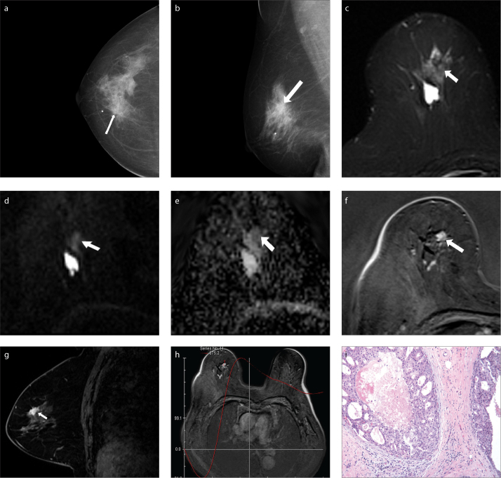
Purpose: We aimed to evaluate the diagnostic accuracy of a combination of dynamic contrast-enhanced magnetic resonance imaging (DCE-MRI) and apparent diffusion coefficient (ADC) values in lesions that manifest with architectural distortion (AD) on mammography.
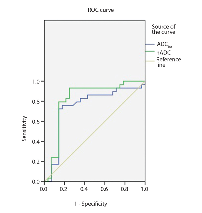
Methods: All full-field digital mammography (FFDM) images obtained between August 2010 and January 2013 were reviewed retrospectively, and 57 lesions showing AD were included in the study. Two independent radiologists reviewed all mammograms and MRI data and recorded lesion characteristics according to the BI-RADS lexicon. The gold standard was histopathologic results from biopsies or surgical excisions and results of the two-year follow-up. Receiver operating characteristic curve analysis was carried out to define the most effective threshold ADC value to differentiate malignant from benign breast lesions. We investigated the sensitivity and specificity of FFDM, DCE-MRI, FFDM+DCE-MRI, and DCE-MRI+ADC.
Results: Of the 57 lesions analyzed, 28 were malignant and 29 were benign. The most effective threshold for the normalized ADC (nADC) was 0.61 with 93.1% sensitivity and 75.0% specificity. The sensitivity and specificity of DCE-MRI combined with nADC was 92.9% and 79.3%, respectively. DCE-MRI combined with nADC showed the highest specificity and equal sensitivity compared with other modalities, independent of the presentation of calcification.
Conclusion: DCE-MRI combined with nADC values was more reliable than mammography in differentiating the nature of disease manifesting as primary AD on mammography.
Figures
References
-
- American College of Radiology. Illustrated breast imaging reporting and data system (BI-RADS): ultrasound. Reston, VA: American College of Radiology; 2003.
-
- Knutzen AM, Gisvold JJ. Likelihood of malignant disease for various categories of mammographically detected, nonpalpable breast lesions. Mayo Clin Proc. 1993;68:454–460. http://dx.doi.org/10.1016/S0025-6196(12)60194-3. - DOI - PubMed
-
- Shaheen R, Schimmelpenninck CA, Stoddart L, Raymond H, Slanetz PJ. Spectrum of diseases presenting as architectural distortion on mammography: multimodality radiologic imaging with pathologic correlation. Semin Ultrasound CT MR. 2011;32:351–362. http://dx.doi.org/10.1053/j.sult.2011.03.008. - DOI - PubMed
-
- Burrell HC, Sibbering DM, Wilson AR, et al. Screening interval breast cancers: mammographic features and prognosis factors. Radiology. 1996;199:811–817. http://dx.doi.org/10.1148/radiology.199.3.8638010. - DOI - PubMed
MeSH terms
Substances
LinkOut - more resources
Full Text Sources
Other Literature Sources
Medical
