. 2016 May-Jul:52-54:127-140.
doi: 10.1016/j.matbio.2016.02.005. Epub 2016 Feb 18.

VEGF stimulates intramembranous bone formation during craniofacial skeletal development

Affiliations

VEGF stimulates intramembranous bone formation during craniofacial skeletal development

Xuchen Duan et al. Matrix Biol. 2016 May-Jul.

Abstract

Deficiency of vascular endothelial growth factor A (VEGF) has been associated with severe craniofacial anomalies in both humans and mice. Cranial neural crest cell (NCC)-derived VEGF regulates proliferation, vascularization and ossification of cartilage and membranous bone. However, the function of VEGF derived from specific subpopulations of NCCs in controlling unique aspects of craniofacial morphogenesis is not clear. In this study a conditional knockdown strategy was used to genetically delete Vegfa expression in Osterix (Osx) and collagen II (Col2)-expressing NCC descendants. No major defects in calvaria and mandibular morphogenesis were observed upon knockdown of VEGF in the Col2(+) cell population. In contrast, loss of VEGF in Osx(+) osteoblast progenitor cells led to reduced ossification of calvarial and mandibular bones without affecting the formation of cartilage templates in newborn mice. The early stages of ossification in the developing jaw revealed decreased initial mineralization levels and a reduced thickness of the collagen I (Col1)-positive bone template upon loss of VEGF in Osx(+) precursors. Increased numbers of proliferating cells were detected within the jaw mesenchyme of mutant embryos. Explant culture assays revealed that mandibular osteogenesis occurred independently of paracrine VEGF action and vascular development. Reduced VEGF expression in mandibles coincided with increased phospho-Smad1/5 (P-Smad1/5) levels and bone morphogenetic protein 2 (Bmp2) expression in the jaw mesenchyme. We conclude that VEGF derived from Osx(+) osteoblast progenitor cells is required for optimal ossification of developing mandibular bones and modulates mechanisms controlling BMP-dependent specification and expansion of the jaw mesenchyme.

Keywords: Bone development; Calvaria; Craniofacial ossification; Mandible; Osterix; VEGF.


Figures

Fig. 1
Fig. 1
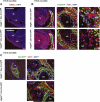
Osx+ and Col2+ progenitor cell populations are located in developing cranial and mandibular regions. (A) The plane of section for which the images in A, B and C are shown is indicated with the dashed line. In situ hybridization on frontal head sections of Vegfa+/+; Osx-Cre:GFP mice at E14.5 (top) and E16.5 (bottom) for Osx (red) and Col2a1 (turquoise) (scale bars: 400 μm). To the right are magnified views of delineated areas of calvaria and mandible in images on left showing Osx and Col2a1 expression (scale bars: 200 μm). c, cartilage primordium of orbito-sphenoid bone; mk, Meckel's cartilage. (B) Analysis of different cell populations located in the head sections of E14.5 and E16.5 mice. Left: head sections from E14.5 (top) and E16.5 (bottom) Vegfa+/+; Osx-Cre:GFP mice indicating Osx/GFP+ cells. Right: head sections from E14.5 (top) and E16.5 (bottom) Vegfa+/+; Tomato; Col2-Cre mice showing cells derived from Col2+ osteochondroprogenitor cells (scale bar: 400 μm). (C) In situ hybridization on frontal head sections of E14.5 Vegfa+/+; Osx-Cre:GFP (top) and Vegfafl/fl; Osx-Cre:GFP (bottom) mice for Osx (red) and Vegfa (turquoise) (scale bars: 200 μm). (For interpretation of the references to color in this figure legend, the reader is referred to the web version of this article.)
Fig. 2
Fig. 2
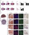
Osx+ osteoblast progenitor cell-derived VEGF regulates intramembranous ossification in developing craniofacial bones. (A) Whole-mount staining with Alizarin Red and Alcian Blue of skulls of P1 Vegfa+/+; Osx-Cre:GFP (top) and Vegfafl/fl; Osx-Cre:GFP (bottom) mice. Sagittal suture width indicated with double headed arrow. p, parietal bone; f, frontal bone; md, mandible; mk, Meckel's cartilage; pal, palatal bone. n = 9/8. Right graphs: Quantification of relative mandibular ossification (n = 8/8), relative mandibular length (n = 9/8), and relative width of sagittal suture (n = 6/6). Values represent mean values ± s.d. P < 0.01 for comparison between genotypes. (B) Top: The plane of section for which the images in B are shown is indicated with the dashed line. Bottom: Histological analysis of frontal head section of P1 Vegfa+/+; Osx-Cre:GFP mice by H&E staining (scale bar: 400 μm). To the right are magnified views of delineated areas of calvaria and mandible in bottom image on left showing Von Kossa staining, anti-Col1 staining, Osx/GFP+ expression, and Trap staining of P1 Vegfa+/+; Osx-Cre:GFP and Vegfafl/fl; Osx-Cre:GFP mice (scale bar: 200 μm). c, cartilage primordium of orbito-sphenoid bone; tb, tooth bud; mk, Meckel's cartilage.
Fig. 3
Fig. 3
VEGF derived from Col2+ cells does not contribute to mineralization in developing craniofacial bones. (A) Whole-mount staining with Alizarin Red and Alcian Blue of skulls of P1 Vegfa+/+; Col2-Cre (top) and Vegfafl/fl; Col2-Cre (bottom) mice. Sagittal suture width indicated with double headed arrow. p, parietal bone; f, frontal bone; md, mandible; mk, Meckel's cartilage; pal, palatal bone. n = 9/9. Right graphs: quantification of relative mandibular ossification (n = 5/8) and relative mandibular length (n = 5/8). Values represent mean values ± s.d. (B) Von Kossa staining of frontal head sections of calvaria and mandibles of P1 Vegfa+/+; Col2-Cre (top) and Vegfafl/fl; Col2-Cre (bottom) mice (scale bar: 200 μm). c, cartilage primordium of orbito-sphenoid bone; tb, tooth bud; mk, Meckel's cartilage.
Fig. 4
Fig. 4
Reduced initial ossification and thickness of the mandibular template upon loss of VEGF in Osx+ precursors. (A) Whole-mount staining with Alizarin Red and Alcian Blue of skulls of E15.5 Vegfa+/+; Osx-Cre:GFP (top) and Vegfafl/fl; Osx-Cre:GFP (bottom) mice. md, mandible; mk, Meckel's cartilage. To the right are magnified views of delineated areas of mandible in images on left. (B) Left: In situ hybridization on frontal head sections of Vegfa+/+; Osx-Cre:GFP (top) and Vegfafl/fl; Osx-Cre:GFP (bottom) mice at E16.5 for Osx (red) and Col2a1 (turquoise) (scale bars: 200 μm). To the right are magnified views of delineated areas of mandible in images on left showing Osx and Col2a1 expression (scale bars: 50 μm). Middle: mandibular sections from E16.5 Vegfa+/+; Osx-Cre:GFP (top) and Vegfafl/fl; Osx-Cre:GFP (bottom) mice indicating Osx/GFP+ cells (scale bars: 200 μm). Mandibular thickness indicated with double headed arrow. Right: Anti-Col1 staining of mandibular sections from E16.5 Vegfa+/+; Osx-Cre:GFP (top) and Vegfafl/fl; Osx-Cre:GFP (bottom) mice (scale bars: 200 μm). tb, tooth bud; mk, Meckel's cartilage. Right graph: quantification of relative mandibular thickness. Values represent mean values ± s.d. P < 0.01 for comparison between genotypes. (For interpretation of the references to color in this figure legend, the reader is referred to the web version of this article.)
Fig. 5
Fig. 5
Loss of VEGF in Osx+ precursors leads to increased cell proliferation without affecting vascular development within jaw mesenchyme. (A) TUNEL staining of mandibular sections from E16.5 Vegfa+/+; Osx-Cre:GFP (top) and Vegfafl/fl; Osx-Cre:GFP (bottom) mice (scale bars: 200 μm). (B) Anti-Ki67 staining of mandibular sections from E16.5 Vegfa+/+; Osx-Cre:GFP (top) and Vegfafl/fl; Osx-Cre:GFP (bottom) mice indicating proliferating cells in tooth bud (left) and jaw mesenchyme (middle) (scale bars: 200 μm). To the right are magnified views of delineated areas of jaw mesenchyme in images on left showing proliferating cells (scale bars: 50 μm). (C) Left: Anti-CD31 staining of mandibular section from E16.5 Vegfa+/+; Osx-Cre:GFP mouse showing Osx/GFP+ cells (scale bar: 200 μm). tb, tooth bud; v, mandibular artery; mk, Meckel's cartilage. Right: magnified views of delineated area of mandible in image on left showing mandibular artery of E16.5 Vegfa+/+; Osx-Cre:GFP (top) and Vegfafl/fl; Osx-Cre:GFP (bottom) mice (scale bar: 200 μm). Arrows indicate CD31 expression in the mandibular artery and blood vessels, asterisk marks blood cells.
Fig. 6
Fig. 6
Mandibular ossification is not stimulated by paracrine VEGF signaling and loss of VEGFR2 in Osx+ precursors does not affect intramembranous bone formation. (A) a. Overview of mandibular dissection procedure on E15.5 embryos. b. Schematic overview of the experimental procedures of the mandibular explant culture. c. Quantification of relative mandibular ossification (n = 5/5). Values represent mean values ± s.d. (B) In situ hybridization on frontal head sections of Vegfr2 +/+; Osx-Cre:GFP mice at E14.5 (top) and E16.5 (bottom) for Osx (red) and Vegfr2 (turquoise) (scale bars: 400 μm). To the right are magnified views of delineated areas of mandible in images on left showing Osx and Vegfr2 expression (scale bars: 200 μm). mk, Meckel's cartilage. (C) Whole-mount Alizarin Red and Alcian Blue-stained skeletal preparations of P1 Vegfr2+/+; Osx-Cre:GFP (top) and Vegfr2fl/fl; Osx-Cre:GFP (bottom) mice. md, mandible; mk, Meckel's cartilage. (For interpretation of the references to color in this figure legend, the reader is referred to the web version of this article.)
Fig. 7
Fig. 7
Reduced VEGF expression coincides with increased BMP signaling within jaw mesenchyme. (A) a. Schematic overview of the experimental procedures of the mandibular explant culture upon knockdown of VEGF. b. Western blotting of GFP and Col1 in whole mandibular lysates from Vegfafl/fl embryos treated with adenoviral Cre compared to adenoviral GFP (control). β-actin is loading control. c. Quantification of relative mandibular ossification (n = 3/3). Values represent mean values ± s.d. d. Western blotting of Ihh, β-catenin, and P-Smad1/5 in whole mandibular lysates from Vegfafl/fl embryos treated with adenoviral Cre compared to adenoviral GFP (control). HSP90 and β-actin are loading control. (B) In situ hybridization on frontal head sections of Vegfa+/+; Osx-Cre:GFP (top) and Vegfafl/fl; Osx-Cre:GFP (bottom) mice at E14.5 for Osx (red) and Bmp2 (turquoise) (scale bars: 200 μm). To the right are magnified views of delineated areas of jaw mesenchyme in images on left showing Osx and Bmp2 expression (scale bars: 100 μm). mk, Meckel's cartilage. (For interpretation of the references to color in this figure legend, the reader is referred to the web version of this article.)
Fig. 8
Fig. 8
Model summarizing the paracrine and autocrine actions of VEGF in the jaw mesenchyme.VEGF produced by Osx+ osteoblast progenitors stimulates mandibular ossification in an autocrine manner. In contrast, NCC-derived VEGF enhances vascular development via paracrine signaling, thereby regulating jaw extension and osteogenesis.

Similar articles

Cited by

References

    1. Bronner ME, LeDouarin NM. Development and evolution of the neural crest: an overview. Dev. Biol. 2012;366:2–9. - PMC - PubMed
    1. Chai Y, Maxson RE., Jr. Recent advances in craniofacial morphogenesis. Dev. Dyn. 2006;235:2353–2375. - PubMed
    1. Weston JA, Thiery JP. Pentimento: neural crest and the origin of mesectoderm. Dev. Biol. 2015;401:37–61. - PubMed
    1. Stalmans I, Lambrechts D, De Smet F, Jansen S, Wang J, Maity S, Kneer P, von der Ohe M, Swillen A, Maes C, Gewillig M, Molin DG, Hellings P, Boetel T, Haardt M, Compernolle V, Dewerchin M, Plaisance S, Vlietinck R, Emanuel B, Gittenberger-de Groot AC, Scambler P, Morrow B, Driscol DA, Moons L, Esguerra CV, Carmeliet G, Behn-Krappa A, Devriendt K, Collen D, Conway SJ, Carmeliet P. VEGF: a modifier of the del22q11 (DiGeorge) syndrome? Nat. Med. 2003;9:173–182. - PubMed
    1. Wiszniak S, Mackenzie FE, Anderson P, Kabbara S, Ruhrberg C, Schwarz Q. Neural crest-derived VEGF promotes embryonic jaw extension. Proc. Natl. Acad. Sci. U. S. A. 2015;112:6086–6091. - PMC - PubMed

Publication types

Substances

LinkOut - more resources