. 2016 May 15:132:79-92.
doi: 10.1016/j.neuroimage.2016.02.032. Epub 2016 Feb 16.

Unmasking local activity within local field potentials (LFPs) by removing distal electrical signals using independent component analysis

Affiliations

Unmasking local activity within local field potentials (LFPs) by removing distal electrical signals using independent component analysis

Nathan W Whitmore et al. Neuroimage. .

Abstract

Local field potentials (LFPs) are commonly thought to reflect the aggregate dynamics in local neural circuits around recording electrodes. However, we show that when LFPs are recorded in awake behaving animals against a distal reference on the skull as commonly practiced, LFPs are significantly contaminated by non-local and non-neural sources arising from the reference electrode and from movement-related noise. In a data set with simultaneously recorded LFPs and electroencephalograms (EEGs) across multiple brain regions while rats perform an auditory oddball task, we used independent component analysis (ICA) to identify signals arising from electrical reference and from volume-conducted noise based on their distributed spatial pattern across multiple electrodes and distinct power spectral features. These sources of distal electrical signals collectively accounted for 23-77% of total variance in unprocessed LFPs, as well as most of the gamma oscillation responses to the target stimulus in EEGs. Gamma oscillation power was concentrated in volume-conducted noise and was tightly coupled with the onset of licking behavior, suggesting a likely origin of muscle activity associated with body movement or orofacial movement. The removal of distal signal contamination also selectively reduced correlations of LFP/EEG signals between distant brain regions but not within the same region. Finally, the removal of contamination from distal electrical signals preserved an event-related potential (ERP) response to auditory stimuli in the frontal cortex and also increased the coupling between the frontal ERP amplitude and neuronal activity in the basal forebrain, supporting the conclusion that removing distal electrical signals unmasked local activity within LFPs. Together, these results highlight the significant contamination of LFPs by distal electrical signals and caution against the straightforward interpretation of unprocessed LFPs. Our results provide a principled approach to identify and remove such contamination to unmask local LFPs.

Keywords: 1/f power spectrum; Basal forebrain; Event-related potential; Functional connectivity; Gamma oscillation; Movement artifact.


Figures

Fig. 1
Fig. 1
Schematic of ICA decomposition and LFP/EEG recording in the auditory oddball task (A) The hypothesis tested in this study specified that LFP signals are consisted of a mix of true local activity (blue) and distal sources, including electrical signals at the reference site (green) and other volume-conducted noise (red). The same color labels are used throughout all figures. (B) Schematic of the ICA analysis. Epochs of LFP and EEG signals around tone onsets were transformed by ICA into the product of a mixing matrix and underlying signal sources called Independent Components (ICs). ICs were classified into the three categories: electrical reference, volume-conducted noise, and local activity. The mixing matrix describes the weights with which ICs are added together to recreate the original signal in each channel. (C) In the auditory oddball task, a standard tone (10 kHz) was presented once every 2 s, and occasionally once every 6–14 s, a deviant oddball tone (6 kHz) was presented that signaled reward if responded to within a 3-s window (yellow). (D) Schematic of the recording configuration. LFPs were simultaneously recorded from a 32-channel linear probe spanning multiple cortical layers in the frontal cortex, and from multi-electrode bundles in bilateral BF. In addition, EEG signals from the frontal cortex and visual cortex were recorded with skull screws. Electrical reference was a skull screw over the cerebellum.
Fig. 2
Fig. 2
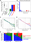
Identification of distal electrical signals originating from electrical reference and volume-conducted noise (A) Left, data from one example session illustrate the identification of the reference signal IC. For each IC, the vector angle between its weights across channels and a vector representing an ideal reference signal with equal weights on all channels was calculated. A single IC representing the electrical signal from the reference site was defined as the IC whose weights had a consistent sign across all channels and from this group the IC with the smallest variation in weight across channels (smallest vector angle, solid green circle). Right, mean distance from the ideal reference for each class of IC across all sessions. (B) Histogram of the peak frequency of non-reference ICs from all sessions reveals a bimodal distribution. ICs with peak frequencies above 45 Hz were identified as volume-conducted noise ICs (red). (C) Log–log plot of the power spectral density of the reference IC from each of the six sessions (green) compared to local ICs (blue, mean ± SEM). The power spectral density of reference ICs deviates from the 1/f power-frequency scaling observed in local ICs, particularly at high frequencies. (D) Log–log plot of the power spectral density of volume-conducted noise ICs (red, mean ± SEM) compared to local ICs (blue, mean ± SEM). The power spectral density of noise ICs also deviates from the 1/f relationship and show a broad peak at 40–60 Hz as well as a narrow peak at 60 Hz corresponding to line noise. (E) The percentage variance accounted for (PVAF) by local activity, electrical reference and volume-conducted noise in each of the six sessions (n = 5 rats), plotted separated for EEG and LFP signals. Sessions were separately sorted for EEGs (left) and LFPs (right) by descending PVAF accounted for by putative local activity ICs.
Fig. 3
Fig. 3
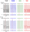
An example of how raw LFP/EEG signals are partitioned into three distinct signal sources (A) Activity from a single oddball trial was partitioned into the sum of local activity, electrical reference and volume-conducted noise signals based on ICA transformation. Removal of distal electrical signals unmasked the dynamics of LFP/EEG signals in single trials. Red vertical lines indicate oddball sound onset. (B) Average LFP/EEG signals of all oddball hit trials from the same session. The main features of event-related responses in both EEGs and LFPs were preserved in the local activity, while volume-conducted noise and electrical reference contributed little to the average response. The mark to the left of each trace indicates zero in the y-axis.
Fig. 4
Fig. 4
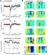
ERP and layer-specific LFP responses are preserved in reconstructed local activity (A) Frontal ERPs from all sessions (colored lines) and the group mean (black), before (top) and after (middle) removing volume-conducted noise and reference ICs. Bottom, pre- and post-correction ERPs were highly similar in the N1-like component window, measured by sliding window cross correlation. Also note that the correction procedure made longer latency ERPs more consistent across sessions and revealed a P3-like component. (B) Comparison of layer-specific frontal LFP responses in oddball hit trials before and after the removal of distal electrical signals in all six sessions. Removing distal electrical signals significantly reduced layer-nonspecific high-frequency noise, while preserving the layer-specific LFP response pattern in the N1-like component window. Sessions were sorted in the same order as the right panel in Fig. 2E. Sessions 1 and 5 were from the same animal.
Fig. 5
Fig. 5
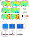
Removal of distal electrical signals affects event-related spectral perturbation (A) Event-related spectral perturbation (ERSP) in oddball hit trials, averaged across all six sessions. Increased gamma oscillation (40–100 Hz) power at long latencies (> 300 ms) was prominent in both the frontal (left) and the visual cortex (right) EEGs prior to the removal of distal electrical signals (top) but was reduced after the correction (middle). Strong long-latency gamma ERSP was observed in the volume-conducted noise (bottom) and was largely eliminated in a frontal-V1 bipolar derivation of the uncorrected data (top right), confirming that the gamma ERSP resulted primarily from signals common to the frontal and V1 EEGs. (B) Mean ERSP in oddball hit trials, averaged across 40–100 Hz between 300 and 1000 ms was significantly reduced after the removal of distal electrical signals (p = 0.021, 2-tailed paired t-test). Most of the gamma range ERSP in the uncorrected EEGs was instead preserved in the noise components. Data from the frontal (solid line) and the visual cortex (dashed line) were combined. Each session was color coded as in Fig. 4A. (C and D) Single-trial gamma oscillation amplitude in the frontal EEG in a representative session (C) and averaged for each session (n = 6) (D), aligned at the first Lick. gamma oscillation amplitude in oddball hit trials showed a stereotypical increase in noise components (middle) but not in local components (left), coincident with the start of licking. This pattern of gamma oscillation increase was similarly present when rats licked outside of the reward window (right, false alarms). Each session in (D) was color coded as in Fig. 4A.
Fig. 6
Fig. 6
Removal of distal electrical signals selectively reduces correlation between distant brain regions Correlation coefficients (mean ± SEM) of un-averaged time series between channel pairs within and between different brain regions, plotted separately for uncorrected (black), corrected (blue), and volume-conducted noise (red) signals. Removal of distal electrical signals significantly decreased the mean correlation of pairs of recordings that spanned two different brain regions but did not decrease correlation between recordings within the same region (2 tailed paired t-test). On the other hand, the aggregate volume-conducted noise was highly correlated across distant brain regions compared to local activity.
Fig. 7
Fig. 7
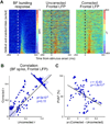
Removal of distal electrical signals improves single-trial amplitude coupling between frontal LFP and BF neuronal activity (A) An example session showing single-trial BF bursting activity (left) and one representative frontal LFP channel, before (middle) and after (right) the removal of distal electrical signals. Oddball and standard trials were pooled and sorted based on BF bursting amplitude in the 50- to 200-ms window. The single-trial coupling between BF bursting amplitude and frontal LFP activity was significantly enhanced after the correction. (B) Scatter plot showing correlation coefficients between single-trial BF bursting strength and frontal LFP amplitude, before (abscissa) and after (ordinate) the removal of distal electrical signals. Each dot represents one frontal LFP channel in one session. LFP channels showing a significant correlation with BF bursting strength (p < 0.0001) are indicated by filled symbols. Histogram along the diagonal line shows a significant increase in correlation coefficients after the removal of distal electrical signals for significantly correlated frontal LFP channels (p < 3 × 10⁻⁶) but not for uncorrelated frontal LFP channels (p = 0.15, 2-tailed paired t-test). (C) In frontal LFP channels that were significantly correlated with BF bursting strength, the change in correlation coefficients induced by the removal of distal electrical signals was negatively correlated with the amount of variance explained by local activity in the uncorrected LFP in each channel.

Similar articles

Cited by

References

    1. Bédard C, Destexhe A. Macroscopic models of local field potentials and the apparent 1/f noise in brain activity. Biophys. J. 2009;96:2589–2603. - PMC - PubMed
    1. Bédard C, Kröger H, Destexhe A. Does the 1/f frequency scaling of brain signals reflect self-organized critical states? Phys. Rev. Lett. 2006;97:118102. - PubMed
    1. Bell AJ, Sejnowski TJ. An information-maximization approach to blind separation and blind deconvolution. Neural Comput. 1995;7:1129–1159. - PubMed
    1. Bosman CA, Schoffelen JM, Brunet N, Oostenveld R, Bastos AM, Womelsdorf T, Rubehn B, Stieglitz T, De Weerd P, Fries P. Attentional stimulus selection through selective synchronization between monkey visual areas. Neuron. 2012;75:875–888. - PMC - PubMed
    1. Buzsáki G, Anastassiou CA, Koch C. The origin of extracellular fields and currents—EEG, ECoG, LFP and spikes. Nat. Rev. Neurosci. 2012;13:407–420. - PMC - PubMed

Publication types