

AAV Gene Therapy for MPS1-associated Corneal Blindness
- PMID: 26899286
- PMCID: PMC4761992
- DOI: 10.1038/srep22131
AAV Gene Therapy for MPS1-associated Corneal Blindness
Abstract
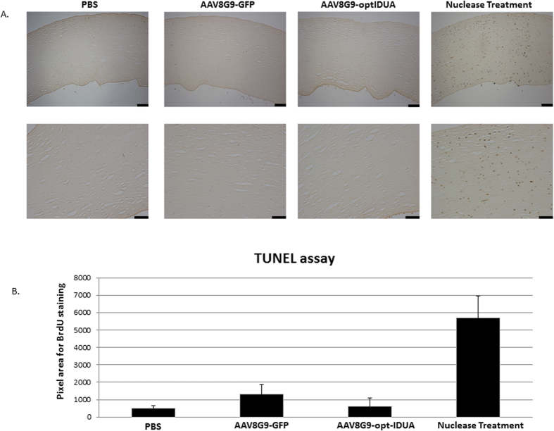
Although cord blood transplantation has significantly extended the lifespan of mucopolysaccharidosis type 1 (MPS1) patients, over 95% manifest cornea clouding with about 50% progressing to blindness. As corneal transplants are met with high rejection rates in MPS1 children, there remains no treatment to prevent blindness or restore vision in MPS1 children. Since MPS1 is caused by mutations in idua, which encodes alpha-L-iduronidase, a gene addition strategy to prevent, and potentially reverse, MPS1-associated corneal blindness was investigated. Initially, a codon optimized idua cDNA expression cassette (opt-IDUA) was validated for IDUA production and function following adeno-associated virus (AAV) vector transduction of MPS1 patient fibroblasts. Then, an AAV serotype evaluation in human cornea explants identified an AAV8 and 9 chimeric capsid (8G9) as most efficient for transduction. AAV8G9-opt-IDUA administered to human corneas via intrastromal injection demonstrated widespread transduction, which included cells that naturally produce IDUA, and resulted in a >10-fold supraphysiological increase in IDUA activity. No significant apoptosis related to AAV vectors or IDUA was observed under any conditions in both human corneas and MPS1 patient fibroblasts. The collective preclinical data demonstrate safe and efficient IDUA delivery to human corneas, which may prevent and potentially reverse MPS1-associated cornea blindness.
Conflict of interest statement
R. Jude Samulski is the founder and a shareholder at Asklepios BioPharmaceutical. He receives research support through the University of North Carolina from Asklepios. BioPharmaceutical. He holds patents that have been licensed by UNC to Asklepios. Biopharmaceutical, for which he receives royalties. He has consulted for Baxter Healthcare and has received payment for speaking. Matthew Hirsch has disclosed AAV8G9-optIDUA to the University of North Carolina.
Figures
Similar articles
-
Mucopolysaccharidoses type I gene therapy.J Inherit Metab Dis. 2021 Sep;44(5):1088-1098. doi: 10.1002/jimd.12414. Epub 2021 Jul 9. J Inherit Metab Dis. 2021. PMID: 34189746 Free PMC article. Review.
-
Intrastromal Gene Therapy Prevents and Reverses Advanced Corneal Clouding in a Canine Model of Mucopolysaccharidosis I.Mol Ther. 2020 Jun 3;28(6):1455-1463. doi: 10.1016/j.ymthe.2020.04.004. Epub 2020 Apr 11. Mol Ther. 2020. PMID: 32330426 Free PMC article.
-
Correction of metabolic, craniofacial, and neurologic abnormalities in MPS I mice treated at birth with adeno-associated virus vector transducing the human alpha-L-iduronidase gene.Mol Ther. 2004 Jun;9(6):866-75. doi: 10.1016/j.ymthe.2004.03.011. Mol Ther. 2004. PMID: 15194053
-
Ocular Tolerability and Immune Response to Corneal Intrastromal AAV-IDUA Gene Therapy in New Zealand White Rabbits.Mol Ther Methods Clin Dev. 2020 May 22;18:24-32. doi: 10.1016/j.omtm.2020.05.014. eCollection 2020 Sep 11. Mol Ther Methods Clin Dev. 2020. PMID: 32542182 Free PMC article.
-
Corneal Regeneration Using Gene Therapy Approaches.Cells. 2023 Apr 28;12(9):1280. doi: 10.3390/cells12091280. Cells. 2023. PMID: 37174680 Free PMC article. Review.
Cited by
-
Adeno-Associated Virus Vector Mobilization, Risk Versus Reality.Hum Gene Ther. 2020 Oct;31(19-20):1054-1067. doi: 10.1089/hum.2020.118. Hum Gene Ther. 2020. PMID: 32829671 Free PMC article.
-
Inhibition of experimental autoimmune uveitis by intravitreal AAV-Equine-IL10 gene therapy.PLoS One. 2022 Aug 18;17(8):e0270972. doi: 10.1371/journal.pone.0270972. eCollection 2022. PLoS One. 2022. PMID: 35980983 Free PMC article.
-
Mucopolysaccharidoses type I gene therapy.J Inherit Metab Dis. 2021 Sep;44(5):1088-1098. doi: 10.1002/jimd.12414. Epub 2021 Jul 9. J Inherit Metab Dis. 2021. PMID: 34189746 Free PMC article. Review.
-
Single stranded adeno-associated virus achieves efficient gene transfer to anterior segment in the mouse eye.PLoS One. 2017 Aug 1;12(8):e0182473. doi: 10.1371/journal.pone.0182473. eCollection 2017. PLoS One. 2017. PMID: 28763501 Free PMC article.
-
Human MiniPromoters for ocular-rAAV expression in ON bipolar, cone, corneal, endothelial, Müller glial, and PAX6 cells.Gene Ther. 2021 Jun;28(6):351-372. doi: 10.1038/s41434-021-00227-z. Epub 2021 Feb 2. Gene Ther. 2021. PMID: 33531684 Free PMC article.
References
-
- Aldenhoven M. et al. Long-term outcome of Hurler syndrome patients after hematopoietic cell transplantation: an international multicenter study. Blood 125(13), p. 2164–72 (2015). - PubMed
-
- Huang Y. et al. Ultrastructural study of the cornea in a bone marrow-transplanted Hurler syndrome patient. Exp Eye Res 62(4), p. 377–87 (1996). - PubMed
Publication types
MeSH terms
Substances
Grants and funding
LinkOut - more resources
Full Text Sources
Other Literature Sources
Medical
Research Materials
Miscellaneous
