. 2016 Feb 22:6:21607.
doi: 10.1038/srep21607.

High-throughput proteomics reveal alarmins as amplifiers of tissue pathology and inflammation after spinal cord injury

Affiliations

High-throughput proteomics reveal alarmins as amplifiers of tissue pathology and inflammation after spinal cord injury

Athanasios Didangelos et al. Sci Rep. .

Abstract

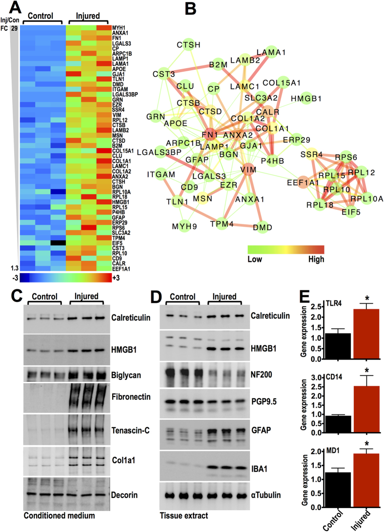
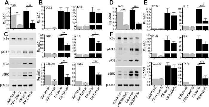
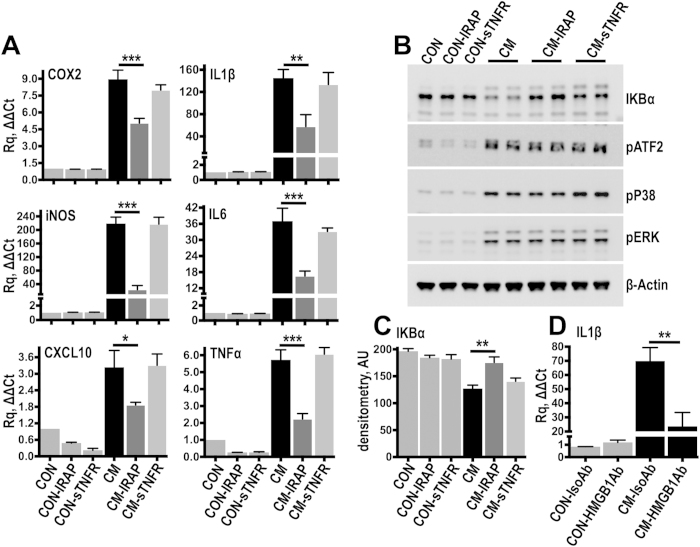
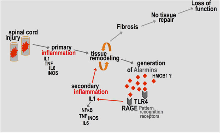
Spinal cord injury is characterized by acute cellular and axonal damage followed by aggressive inflammation and pathological tissue remodelling. The biological mediators underlying these processes are still largely unknown. Here we apply an innovative proteomics approach targeting the enriched extracellular proteome after spinal cord injury for the first time. Proteomics revealed multiple matrix proteins not previously associated with injured spinal tissue, including small proteoglycans involved in cell-matrix adhesion and collagen fibrillogenesis. Network analysis of transcriptomics and proteomics datasets uncovered persistent overexpression of extracellular alarmins that can trigger inflammation via pattern recognition receptors. In mechanistic experiments, inhibition of toll-like receptor-4 (TLR4) and the receptor for advanced glycation end-products (RAGE) revealed the involvement of alarmins in inflammatory gene expression, which was found to be dominated by IL1 and NFκΒ signalling. Extracellular high-mobility group box-1 (HMGB1) was identified as the likely endogenous regulator of IL1 expression after injury. These data reveal a novel tissue remodelling signature and identify endogenous alarmins as amplifiers of the inflammatory response that promotes tissue pathology and impedes neuronal repair after spinal cord injury.


Figures

Figure 1
Figure 1. Proteomics analysis of ECM-enriched protein fraction of injured spinal tissue.
(A,B) Protein composition in 0.08% SDS and 4 M guanidine extracts derived from T10 injury epicentre spinal cord explants, 8 weeks post-contusion. Ponceau staining (A) shows a clear difference in the protein content of the 2 extracts. 15 μg of protein was loaded in both lanes. Immunoblotting (B) depicts the relative abundance of selected proteins in SDS and guanidine extracts. (C) Average number of normalized spectral counts measured in 4 M guanidine extracts of 3 uninjured control and 3 injured T10 spinal cord specimens by shot-gun LC-MS/MS with a total of 2346 proteins identified, 10 ppM peptide mass accuracy tolerance, 1% false discovery rate and 95% peptide and protein identification probability. (D) Volcano plot shows differential protein expression measured by spectral counting in 3 uninjured control and 3 injured T10 spinal cord extracts. Proteins are separated according to their spectral count fold-change (Injured/Control; x-axis) and their two-tailed t-test P value (y-axis). P=0.05 and P=0.01 are indicated. Selected highly dysregulated proteins are highlighted (upregulated: red; downregulated: green). (E) Principal component analysis (PCA) clustering control and injured T10 spinal cord extracts based on spectral counting of the 2346 LC-MS/MS protein identifications.
Figure 2
Figure 2. Network analysis of differentially regulated proteins in chronic SCI tissue.
(A,B) The 8 most populated subnetworks generated by MCL clustering (1.9 inflation value & 0.4 edge weight cut-off; StringDB, v9.1) of upregulated (A) and downregulated (B) protein networks shown in Supplementary Figure S1. Individual subnetworks were analysed by BiNGO to identify the predominant gene ontology term. Numbers in parenthesis indicate the number of proteins in the predominant gene ontology versus the total number of proteins in each subnetwork. Node color indicates betweeness centrality while edge color indicates interaction score based on the predicted functional links between nodes (green: low values; red: high values).
Figure 3
Figure 3. Extracellular matrix proteins identified by proteomics and validated by immunoblotting.
(A) Differential expression of 47 typical extracellular matrix proteins identified by LC-MS/MS in uninjured control and injured T10 spinal cords. (B) Relative matrix protein abundance in the guanidine extracts was calculated as the ratio of protein spectral counts divided by protein molecular mass in kDa. Galectin-1 had the highest spectral count/molecular mass ratio (2.2) while collagen alpha-2(IV) (Col4a2) had the lowest ratio (0.01). (C) Different matrix proteins were validated by western-blotting in 4 M guanidine extracts of uninjured control and injured T10 spinal cord specimens, 8 weeks post-contusion. One representative example is shown. Ponceau stain demonstrates comparable loading but distinct protein composition in control versus injured extracts.
Figure 4
Figure 4. Identification of persistently upregulated bioactive molecules.
(A) Heat-map displays 48 upregulated proteins found in proteomics analysis of guanidine extracts 8 weeks post-injury. Proteins are listed on the heat-map according to their fold change (FC, Injured/Controls). These molecules were also upregulated at the transcript level 5 weeks post-injury as detected by microarray analysis (see reference 29). (B) Network analysis (StringDB, v9.1) of the 48 genes upregulated both at the mRNA and protein level. Node color indicates betweeness centrality while edge color indicates interaction score based on the predicted functional links between nodes (green: low values; red: high values). (C,D) Immunoblotting of conditioned medium (C) and tissue extracts (D) derived from either uninjured control or injured T10 spinal cord explants cultured for 24 hours in plain DMEM culture medium. The endogenous TLR4 ligands HMGB1, biglycan, fibronectin (EDA fragments) and tenascin-C are present in the conditioned medium. Calreticulin and HMGB1 were also upregulated in tissue extracts. (E) Relative mRNA expression of TLR4, CD14 and MD1 measured by TaqMan quantitative PCR in either control or injured T10 spinal cord explants, 7 days post-injury. ACTB served as the housekeeping gene. N = 3 animals per group; * P0.05, two-tailed t-test.
Figure 5
Figure 5. TLR4 and RAGE siRNA knockdown partially suppress inflammatory gene expression.
(A,B) TLR4 expression was blocked using siRNA in primary fibroblasts (A). For comparison, control cells were transfected with target-less scrambled (Scr) siRNA. 48 hours later, Scr or TLR4-transfected fibroblasts were stimulated for 3 hours with conditioned medium (CM) sampled from injured spinal cord explants or kept in plain culture medium (CON). Gene expression of inflammatory genes was measured by TaqMan qPCR (B). ACTB served as the housekeeping gene. N = 6 independent experiments; *P0.05, **P0.01, ***P0.001; Anova with Fisher’s LSD multiple comparison test. (C) To examine acute signalling activation, scrambled or TLR4 siRNA transfected cells were stimulated with conditioned medium for 25 minutes. Signalling activation was examined by immunoblotting. N = 3 independent experiments. (D,E) As for TLR4, RAGE expression was blocked using siRNA (D) and 48 hours later Scr or RAGE-transfected fibroblasts were stimulated with conditioned medium (CM) and gene expression was measured by TaqMan qPCR (E). ACTB served as the housekeeping gene. N = 6 independent experiments. ***P0.001; Anova with Fisher’s LSD multiple comparison test. (F) Acute signalling activation of RAGE-transfected cells was examined by immunoblotting. N = 3 independent experiments.
Figure 6
Figure 6. Interleukin 1 is the dominant inflammatory factor in the injury conditioned medium.
(A) Resting fibroblasts were stimulated for 3 hours with injury conditioned medium (CM) supplemented with 20 ng/ml of either IRAP or sTNFR. Control cells were kept in plain culture medium (CON) and additional controls were incubated with IRAP (CON-IRAP) or sTNFR (CON-sTNFR). Gene expression was measured by TaqMan qPCR. ACTB served as the housekeeping gene. N = 4 independent experiments. (B,C) To examine acute signalling activation cells were stimulated for 25 minutes with conditioned medium supplemented either with IRAP or sTNFR and signalling activation was examined by immunoblotting (B). IκΒα levels were quantified by densitometry (C) N = 3 independent experiments. (D) Resting cells were stimulated with injury conditioned medium which was previously treated for 2 hours with HMGB1 antibodies (HMGB1Ab; 5 μg/ml) to neutralize soluble extracellular HMGB1, or with isotype control antibodies (IsoAb; 5 μg/ml). IL1β expression was measured by TaqMan qPCR. N = 6 independent experiments. *P0.05, **P0.01, ***P0.001; Anova with Fisher’s LSD multiple comparison test.
Figure 7
Figure 7. Tissue remodeling and the generation of alarmins after SCI.
Schematic summary of the involvement of danger-associated molecular patterns (DAMPs or alarmins) in spinal cord inflammation. After injury, primary inflammation is driven by proinflammatory cytokines and leads to excessive tissue remodelling and fibrosis. Soluble alarmins are generated during pathological tissue remodelling. They activate pattern recognition receptors and contribute to the persistent (secondary) inflammatory activation in the injury epicentre.

References

    1. Benowitz L. I. & Popovich P. G. Inflammation and axon regeneration. Curr Opin Neurol 24(6), 577–583 (2011). - PubMed
    1. Silver J. & Miller J. H. Regeneration beyond the glial scar. Nat Rev Neurosci 5(2), 146–156 (2004). - PubMed
    1. Schwab M. E. & Strittmatter S. M. Nogo limits neural plasticity and recovery from injury. Curr Opin Neurobiol 27, 53–60 (2014). - PMC - PubMed
    1. Fawcett J. W. & Asher R. A. The glial scar and central nervous system repair. Brain Res Bull 49(6), 377–391 (1999). - PubMed
    1. Alilain W. J., Horn K. P., Hu H., Dick T. E. & Silver J. Functional regeneration of respiratory pathways after spinal cord injury. Nature 475(7355), 196–200 (2011). - PMC - PubMed

Publication types

MeSH terms