. 2016 Feb 22:6:21878.
doi: 10.1038/srep21878.

Replication and shedding of MERS-CoV in Jamaican fruit bats (Artibeus jamaicensis)

Affiliations

Replication and shedding of MERS-CoV in Jamaican fruit bats (Artibeus jamaicensis)

Vincent J Munster et al. Sci Rep. .

Abstract

The emergence of Middle East respiratory syndrome coronavirus (MERS-CoV) highlights the zoonotic potential of Betacoronaviruses. Investigations into the origin of MERS-CoV have focused on two potential reservoirs: bats and camels. Here, we investigated the role of bats as a potential reservoir for MERS-CoV. In vitro, the MERS-CoV spike glycoprotein interacted with Jamaican fruit bat (Artibeus jamaicensis) dipeptidyl peptidase 4 (DPP4) receptor and MERS-CoV replicated efficiently in Jamaican fruit bat cells, suggesting there is no restriction at the receptor or cellular level for MERS-CoV. To shed light on the intrinsic host-virus relationship, we inoculated 10 Jamaican fruit bats with MERS-CoV. Although all bats showed evidence of infection, none of the bats showed clinical signs of disease. Virus shedding was detected in the respiratory and intestinal tract for up to 9 days. MERS-CoV replicated transiently in the respiratory and, to a lesser extent, the intestinal tracts and internal organs; with limited histopathological changes observed only in the lungs. Analysis of the innate gene expression in the lungs showed a moderate, transient induction of expression. Our results indicate that MERS-CoV maintains the ability to replicate in bats without clinical signs of disease, supporting the general hypothesis of bats as ancestral reservoirs for MERS-CoV.


Conflict of interest statement

The authors declare no competing financial interests.

Figures

Figure 1
Figure 1
Replication kinetics of MERS-CoV in cells expressing bat DPP4. (A) Hamster BHK transfected to express Jamaican fruit bat (red) and hamster DPP4 (blue) and inoculated with MERS-CoV (MOI of 1 TCID50/cell). (B) Jamaican fruit bat primary kidney (red) and VeroE6 (blue) cell lines inoculated with MERS-CoV (MOI of 0.01 TCID50/cell). Supernatants were harvested at 0, 24, 48 and 72 hpi and viral titers were determined by end-point titration in quadruplicate in VeroE6 cells. Geometric mean titers were calculated from three independent experiments. Error bars indicate standard deviations.
Figure 2
Figure 2
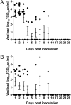
MERS-CoV shedding in Jamaican fruit bats. (A) from the respiratory tract of Jamaican fruit bats. (B) from the intestinal tract of Jamaican fruit bats. Oral and rectal swabs were collected at exam and necropsy dates. RNA was extracted and qRT-PCR was performed. Viral load was expressed as TCID50 equivalents.
Figure 3
Figure 3
MERS-CoV viral load in tissues collected from Jamaican fruit bats inoculated with MERS-CoV. Tissues were collected at necropsy on the indicated days post-inoculation. RNA was extracted and qRT-PCR was performed. Viral load was expressed as TCID50 equivalents per gram tissue.
Figure 4
Figure 4
Histopathological changes in Jamaican fruit bats inoculated with MERS-CoV. Jamaican fruit bats were euthanized on day 2, 4, 7, 14 and 28 days post inoculation and tissue was collected, formalin fixed and stained with hematoxylin and eosin (H&E; panels A,C) or immunohistochemistry using a polyclonal α-MERS-CoV antibody (IHC; panels B,D). Shown are bat 1 (panel A–C) and bat 5 (panel D–F). (A) Bat 1, no pulmonary pathology detected on 2 dpi visible is apparently normal lungtissue. (B) IHC staining of sequential section of panel A reveals abundance of MERS-CoV antigen in bat 1. (C) ISH staining of sequential section of panel (A) reveals abundance of MERS-CoV RNA in bat 1. (D) Bat 5, minimal alveolar thickening with infiltration of small numbers of foamy macrophages and fewer neutrophils (indicated by black arrow). (E) IHC staining of sequential section of panel C reveals abundance of MERS-CoV antigen in affected areas in bat 5. (F) ISH staining of sequential section of panel A reveals abundance of MERS-CoV RNA in bat 5. Original magnification: 20× for (A, B, D) and (E) and 40× for (C) and (F).
Figure 5
Figure 5
MERS-CoV replicates in alveolar pneumocytes. Lungs of Jamaican fruit bat 5 were stained with α-cytokeratin as an epithelial marker (purple) and with a polyclonal α-coronavirus antibody (brown-red) to demonstrate that viral antigen was located along the basement membrane of alveolar pneumocytes of bat 1 at 2 dpi (indicated by black arrows). Original magnification: 40×.
Figure 6
Figure 6
Mx1, ISG56 and RANTES upregulation after MERS-CoV infection in Jamaican fruit bats. Gene expression was determined by qRT-PCR and normalized using HPRT expression. Data are the mean and standard error of mean calculated from two Jamaican fruit bats at each time point and is represented as fold increase over uninfected controls. Statistical significance was calculated using two-tailed unpaired t-tests.

Similar articles

Cited by

References

    1. Zaki A, van Boheemen S, Bestebroer T, Osterhaus A, Fouchier R. Isolation of a novel coronavirus from a man with pneumonia in Saudi Arabia. N Engl J Med. 2012;367:1814–1820. doi: 10.1056/NEJMoa1211721. - DOI - PubMed
    1. WHO. Middle East respiratory syndrome coronavirus (MERS-CoV), http://www.who.int/emergencies/mers-cov/en/ (Date of access: 26/01/2016).
    1. Cotten M, et al. Full-genome deep sequencing and phylogenetic analysis of novel human betacoronavirus. Emerg Infect Dis. 2013;19:736. doi: 10.3201/eid1905.130057. - DOI - PMC - PubMed
    1. van Boheemen, S. et al. Genomic characterization of a newly discovered coronavirus associated with acute respiratory distress syndrome in humans. mBio 3, 10.1128/mBio.00473-12 (2012). - PMC - PubMed
    1. de Groot, R. In Virus Taxonomy (eds Andrew M. Q. King, Michael J. Adams, Eric B. Carstens & Elliot J. Lefkowitz ) 806–828 (Elsevier, 2012).

Publication types