

A ventral salience network in the macaque brain
- PMID: 26899785
- PMCID: PMC4851897
- DOI: 10.1016/j.neuroimage.2016.02.029
A ventral salience network in the macaque brain
Abstract
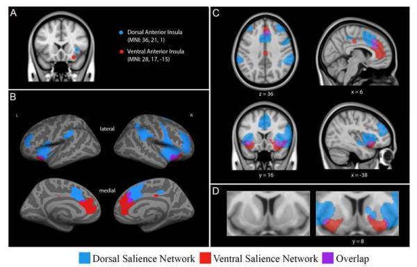
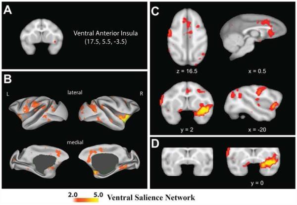
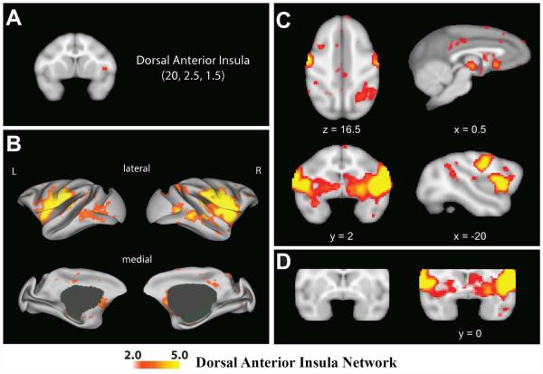
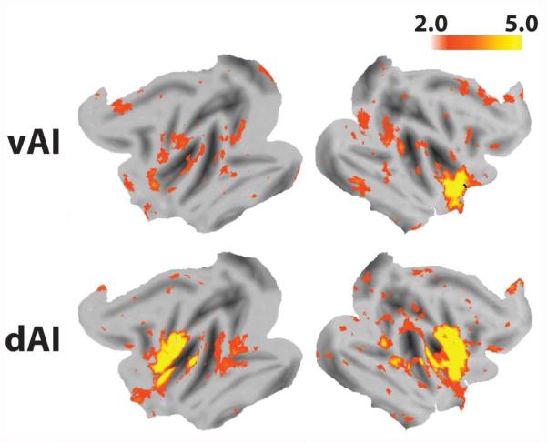
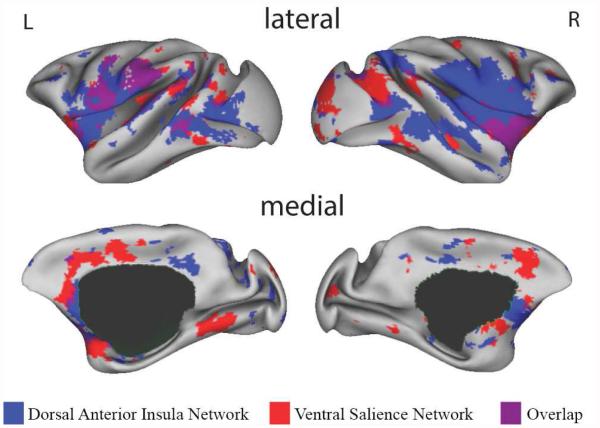
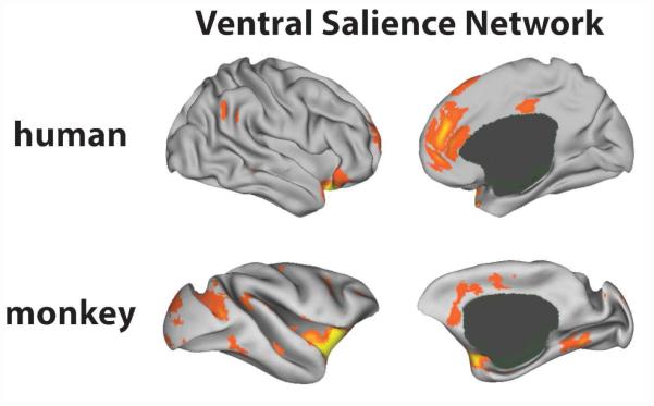
Successful navigation of the environment requires attending and responding efficiently to objects and conspecifics with the potential to benefit or harm (i.e., that have value). In humans, this function is subserved by a distributed large-scale neural network called the "salience network". We have recently demonstrated that there are two anatomically and functionally dissociable salience networks anchored in the dorsal and ventral portions of the human anterior insula (Touroutoglou et al., 2012). In this paper, we test the hypothesis that these two subnetworks exist in rhesus macaques (Macaca mulatta). We provide evidence that a homologous ventral salience network exists in macaques, but that the connectivity of the dorsal anterior insula in macaques is not sufficiently developed as a dorsal salience network. The evolutionary implications of these finding are considered.
Keywords: Intrinsic functional connectivity; Resting state fMRI; Rhesus macaques; Salience network.
Copyright © 2016 Elsevier Inc. All rights reserved.
Figures
Similar articles
-
Salience network connectivity in the insula is associated with individual differences in interoceptive accuracy.Brain Struct Funct. 2017 May;222(4):1635-1644. doi: 10.1007/s00429-016-1297-7. Epub 2016 Aug 29. Brain Struct Funct. 2017. PMID: 27573028
-
Lateralization in intrinsic functional connectivity of the temporoparietal junction with salience- and attention-related brain networks.J Neurophysiol. 2012 Dec;108(12):3382-92. doi: 10.1152/jn.00674.2012. Epub 2012 Sep 26. J Neurophysiol. 2012. PMID: 23019004
-
Altered intrinsic organisation of brain networks implicated in attentional processes in adult attention-deficit/hyperactivity disorder: a resting-state study of attention, default mode and salience network connectivity.Eur Arch Psychiatry Clin Neurosci. 2016 Jun;266(4):349-57. doi: 10.1007/s00406-015-0630-0. Epub 2015 Aug 11. Eur Arch Psychiatry Clin Neurosci. 2016. PMID: 26260900
-
Role of the anterior insula in task-level control and focal attention.Brain Struct Funct. 2010 Jun;214(5-6):669-80. doi: 10.1007/s00429-010-0260-2. Epub 2010 May 29. Brain Struct Funct. 2010. PMID: 20512372 Free PMC article. Review.
-
Social attention and the brain.Curr Biol. 2009 Nov 3;19(20):R958-62. doi: 10.1016/j.cub.2009.08.010. Curr Biol. 2009. PMID: 19889376 Free PMC article. Review.
Cited by
-
Interconnected sub-networks of the macaque monkey gustatory connectome.Front Neurosci. 2023 Feb 16;16:818800. doi: 10.3389/fnins.2022.818800. eCollection 2022. Front Neurosci. 2023. PMID: 36874640 Free PMC article.
-
Group Cognitive Behavior Therapy Reversed Insula Subregions Functional Connectivity in Asthmatic Patients.Front Aging Neurosci. 2017 Apr 18;9:105. doi: 10.3389/fnagi.2017.00105. eCollection 2017. Front Aging Neurosci. 2017. PMID: 28458637 Free PMC article.
-
Connecting Circuits with Networks in Addiction Neuroscience: A Salience Network Perspective.Int J Mol Sci. 2023 May 22;24(10):9083. doi: 10.3390/ijms24109083. Int J Mol Sci. 2023. PMID: 37240428 Free PMC article. Review.
-
Anterior Cingulate Cortex Ablation Disrupts Affective Vigor and Vigilance.J Neurosci. 2021 Sep 22;41(38):8075-8087. doi: 10.1523/JNEUROSCI.0673-21.2021. Epub 2021 Aug 11. J Neurosci. 2021. PMID: 34380767 Free PMC article.
-
Reorganization in the macaque interoceptive-allostatic network following anterior cingulate cortex damage.Cereb Cortex. 2023 Apr 4;33(8):4334-4349. doi: 10.1093/cercor/bhac346. Cereb Cortex. 2023. PMID: 36066407 Free PMC article.
References
-
- Allman J, Hakeem A, Watson K. Two Phylogenetic Specializations in the Human Brain. The Neuroscientist. 2002;8:335–346. - PubMed
Publication types
MeSH terms
Grants and funding
LinkOut - more resources
Full Text Sources
Other Literature Sources