Review
. 2016 Jan 24:22:61-72.
eCollection 2016.

Effects of blue light on the circadian system and eye physiology

Affiliations
Review

Effects of blue light on the circadian system and eye physiology

Gianluca Tosini et al. Mol Vis. .

Abstract

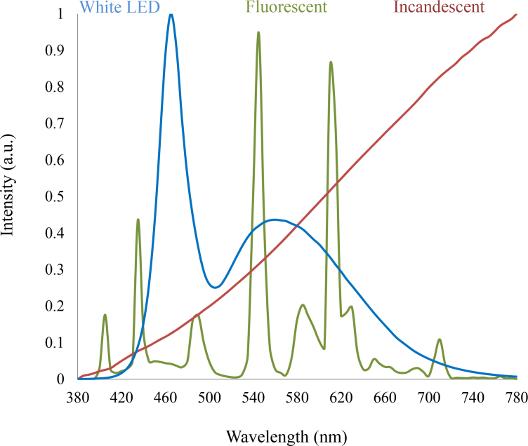
Light-emitting diodes (LEDs) have been used to provide illumination in industrial and commercial environments. LEDs are also used in TVs, computers, smart phones, and tablets. Although the light emitted by most LEDs appears white, LEDs have peak emission in the blue light range (400-490 nm). The accumulating experimental evidence has indicated that exposure to blue light can affect many physiologic functions, and it can be used to treat circadian and sleep dysfunctions. However, blue light can also induce photoreceptor damage. Thus, it is important to consider the spectral output of LED-based light sources to minimize the danger that may be associated with blue light exposure. In this review, we summarize the current knowledge of the effects of blue light on the regulation of physiologic functions and the possible effects of blue light exposure on ocular health.


Figures

Figure 1
Figure 1
A comparison of the power spectrum of a standard white-light LED, a tricolor fluorescent lamp, and an incandescent source. The radically different power spectrums can look similar when viewed directly by the eye, irrespective of how much blue emission is present.
Figure 2
Figure 2
In addition to the classical photoreceptors (rods and cones), ipRGCs are present in the retina. Recent studies have shown that at least two types of intrinsically photosensitive retinal ganglion cells (ipRGCs) have been identified: M1 and M2. Most of the M1 cells project to the suprachiasmatic nucleus (SCN) of the hypothalamus whereas the number of M1 and M2 projecting to the olivary pretectal nucleus (OPN) is similar (55% from M1 cells versus 45% from M2 cells). The M1 cells are considerably smaller but respond with significantly larger depolarizations and light-induced currents than do the M2 cells. Other neural targets of ipRGCs not shown in the figure include the preoptic area, sub-paraventricular zone, anterior hypothalamic nucleus, lateral hypothalamus, medial amygdaloid nucleus, lateral habenula, lateral geniculate nucleus (dorsal division), bed nucleus of the stria terminalis, periaqueductal gray, and superior colliculus. OS=outer segments; IS=inner segments; ONL=outer nuclear layer; OPL=outer plexiform layer; INL=inner nuclear layer; IPL=inner plexiform layer; GCL=ganglion cell layer; from [31] with permission.
Figure 3
Figure 3
Top panels. The exposure to blue light (λmax 474), green light (λmax 513), or fluorescent light at the intensity of 1×10⁻¹ μW/cm2 for 4 h/day for 30 days did not produce a significant change in the number of cells in the photoreceptor layers of the Sprague-Dawley rats (n=6; see [121] for details about the methods used to quantify cells in the photoreceptor layer). Lower panels. The exposure to blue or green light-emitting diodes (LEDs) for 4 h in the middle of the day did not induce apoptosis. Terminal deoxynucleotidyl transferase-mediated uridine 5′-triphosphate-biotin nick end labeling (TUNEL) assay: 4- to 6-week-old Sprague-Dawley rats (n=6) were anesthetized (75 mg/kg ketamine and 23 mg/kg xylazine), kept on heating pads (37 °C), and exposed to blue or green light for 4 h. The pupils were dilated with 1% atropine and 2.5% phenylephrine eye drops 45 min before the light exposure. Rats were then killed 16 h after the exposure to blue light or green light. The eyes were explanted and fixed using freshly prepared 4% polyformaldehyde in PBS, pH 7.4 for 20 min at room temperature. They were washed 3X with PBS, permeabilized with freshly prepared 0.1% Triton X-100 in 0.1% sodium citrate for 2 min on ice (2–8 °C), and then the TUNEL reaction was performed according to the instructions included in the manual (In Situ cell Death Detection kit). The slides were incubated in a humidified container for 60 min at 37 °C in the dark. Slides were rinsed 3X with PBS, and samples were analyzed under a fluorescence microscope (Zeiss Axioskop).
Figure 4
Figure 4
Different light treatments did not affect rhodopsin mRNA levels (one-way ANOVA, p>0.1). Exposure to blue light (λmax 474) at the intensity of 1×10⁻¹ μW/cm2 for 4 h/day for 30 days produced significant changes in the mRNA levels of short wavelength sensitive (SW) opsin, melanopsin, and medium wavelength sensitive opsin (* one-way ANOVA followed by Holm-Sidak tests, p<0.05). Rats were exposed to blue, green, or white light-emitting diodes (LEDs) every day (4 h) for 30 days in the middle of the day (11:00 to 15:00) and then returned to a 12 h:12 h light-dark cycle. The intensity of the light during the light phase of the 12 h:12 h light-dark cycle was about 400–450 lux. Every day, the pupils were dilated with 1% atropine and 2.5% phenylephrine eye drops 45 min before exposure to blue, green, or white light-emitting diodes (LEDs). After 30 days, the rats were killed, and the retinas were explanted, immediately frozen, and stored at −80 °C. mRNA was then extracted, and mRNA levels were measured using real-time quantitative PCR (qPCR; see [122] for details about primers and qPCR conditions).

References

    1. Ferguson I, Melton A, Xu T, Jamil M, Fenwick W. What would Edison do with solid state lighting? Proc. SPIE 7784, Tenth International Conference on Solid State Lighting 2010; 77840A.
    1. Pimputkar S, Speck J, DenBaars S, Nakamura S. Prospects for LED lighting. Nat Photonics. 2009;3:180–2.
    1. Schubert F. Light-Emitting Diodes. Cambridge University Press; 2006; pp. 434.
    1. Nakamura S, Chichibu S. Introduction to Nitride Semiconductor Blue Lasers and Light Emitting Diodes. 2000; CRC Press; 1st 386 pages.
    1. Nakamura S. Present performance of InGaN-based blue/green/yellow LEDs. Light-Emitting Diodes: Research, Manufacturing, and Applications. Proc SPIE. 1997;xxx:26.

Publication types

MeSH terms