

Scleral fibroblast response to experimental glaucoma in mice
- PMID: 26900327
- PMCID: PMC4734151
Scleral fibroblast response to experimental glaucoma in mice
Abstract
Purpose: To study the detailed cellular and molecular changes in the mouse sclera subjected to experimental glaucoma.
Methods: Three strains of mice underwent experimental bead-injection glaucoma and were euthanized at 3 days and 1, 3, and 6 weeks. Scleral protein expression was analyzed with liquid chromatography coupled with tandem mass spectrometry (LC-MS/MS) using (16)O/(18)O labeling for quantification in 1- and 6-week tissues. Sclera protein samples were also analyzed with immunoblotting with specific antibodies to selected proteins. The proportion of proliferating scleral fibroblasts was quantified with Ki67 and 4',6-diamidino-2-phenylindole (DAPI) labeling, and selected proteins were studied with immunohistochemistry.
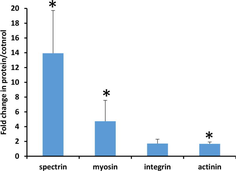
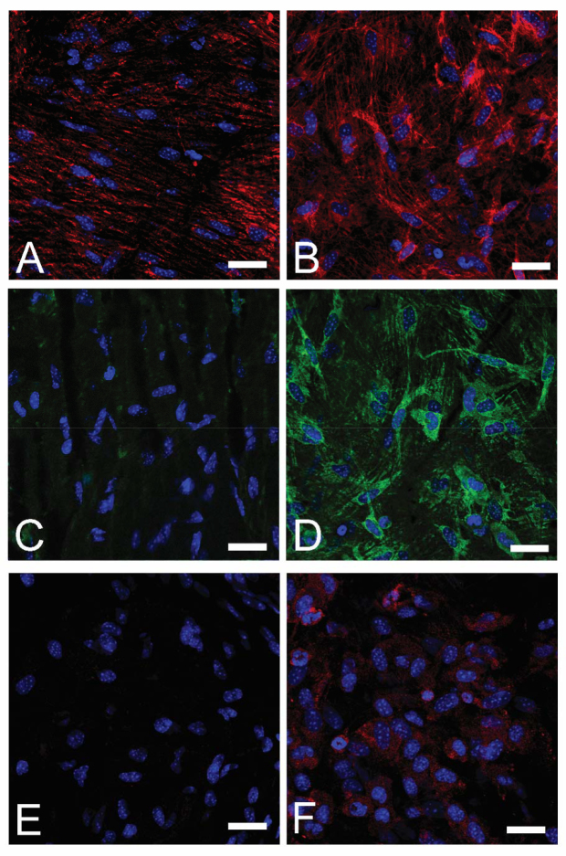
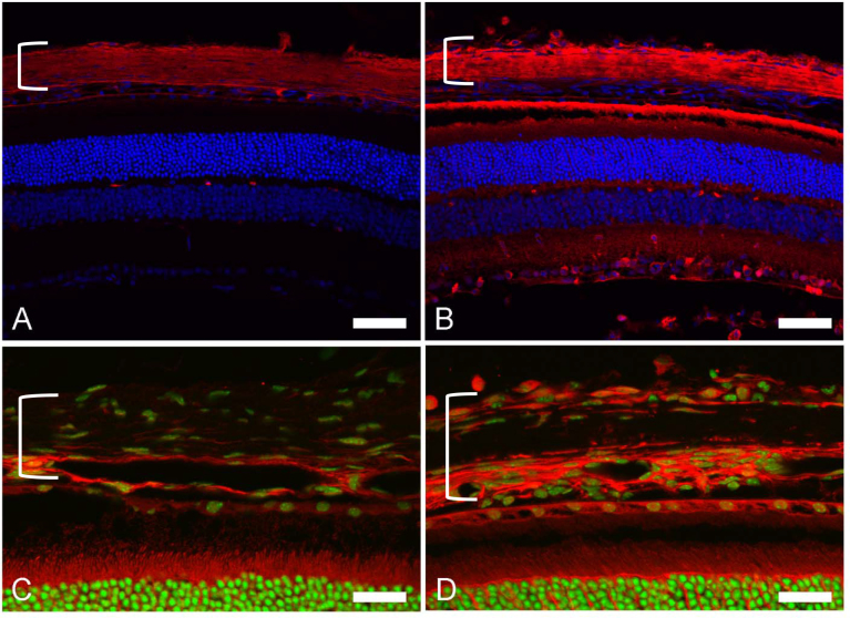
Results: Proteomic analysis showed increases in molecules involved in integrin-linked kinase signaling and actin cytoskeleton signaling pathways at 1 and 6 weeks after experimental glaucoma. The peripapillary scleral region had more fibroblasts than equatorial sclera (p=0.001, n=217, multivariable regression models). There was a sixfold increase in proliferating fibroblasts in the experimental glaucoma sclera at 1 week and a threefold rise at 3 and 6 weeks (p=0.0005, univariate regression). Immunoblots confirmed increases for myosin, spectrin, and actinin at 1 week after glaucoma. Thrombospondin-1 (TSP-1), HINT1, vimentin, actinin, and α-smooth muscle actin were increased according to immunohistochemistry.
Conclusions: Scleral fibroblasts in experimental mouse glaucoma show increases in actin cytoskeleton and integrin-related signaling, increases in cell division, and features compatible with myofibroblast transition.
Figures
References
-
- Burgoyne CF, Downs JC, Bellezza AJ, Suh JK, Hart RT. The optic nerve head as a biomechanical structure: a new paradigm for understanding the role of IOP-related stress and strain in the pathophysiology of glaucomatous optic nerve head damage. Prog Retin Eye Res. 2005;24:39–73. http://www.ncbi.nlm.nih.gov/entrez/query.fcgi?cmd=Retrieve&db=PubMed&lis... - PubMed
-
- Sigal IA, Yang H, Roberts MD, Burgoyne CF, Downs JC. IOP-induced lamina cribrosa displacement and scleral canal expansion: an analysis of factor interactions using parameterized eye-specific models. Invest Ophthalmol Vis Sci. 2011;52:1896–907. http://www.ncbi.nlm.nih.gov/entrez/query.fcgi?cmd=Retrieve&db=PubMed&lis... - PMC - PubMed
-
- Sigal IA, Flanagan JG, Ethier CR. Factors influencing optic nerve head biomechanics. Invest Ophthalmol Vis Sci. 2005;46:4189–99. http://www.ncbi.nlm.nih.gov/entrez/query.fcgi?cmd=Retrieve&db=PubMed&lis... - PubMed
-
- Quigley HA, Addicks EM, Green WR, Maumenee AE. Optic nerve damage in human glaucoma. II. The site of injury and susceptibility to damage. Arch Ophthalmol. 1981;99:635–49. http://www.ncbi.nlm.nih.gov/entrez/query.fcgi?cmd=Retrieve&db=PubMed&lis... - PubMed
-
- Silver DM, Geyer O. Pressure-volume relation for the living human eye. Curr Eye Res. 2000;20:115–20. http://www.ncbi.nlm.nih.gov/entrez/query.fcgi?cmd=Retrieve&db=PubMed&lis... - PubMed
Publication types
MeSH terms
Substances
Grants and funding
LinkOut - more resources
Full Text Sources
Medical
Miscellaneous
