

"Central vessel sign" on 3T FLAIR* MRI for the differentiation of multiple sclerosis from migraine
- PMID: 26900578
- PMCID: PMC4748312
- DOI: 10.1002/acn3.273
"Central vessel sign" on 3T FLAIR* MRI for the differentiation of multiple sclerosis from migraine
Abstract
Objective: The diagnosis of multiple sclerosis (MS) presently relies on radiographic assessments of imperfect specificity. Recent data using T2* methodology for the detection of the "central vessel sign" (CVS) in MS lesions suggests this novel MRI technique may distinguish MS from other disorders. Our aim was to determine if evaluation for CVS on 3T FLAIR* MRI differentiates MS from migraine.
Methods: Patients with MS or migraine and a prior brain MRI demonstrating at least two hyperintense lesions ≥3 mm were recruited. Exclusion criteria included any additional comorbidity known to cause brain MRI abnormalities. 3T MRI was performed in each participant with administration of gadopentetate dimeglumine, and FLAIR* images were generated in postprocessing. The total number of discrete ovoid lesions ≥3 mm were counted on FLAIR, per participant, and subsequently evaluated for presence of CVS on FLAIR*. An exploratory method evaluating for CVS in a maximum of 12 lesions per subject was also completed.
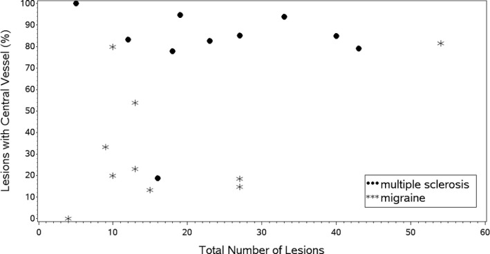
Results: Ten participants with MS and 10 with migraine completed the study. The median percentage (quartiles) of lesions in MS participants with CVS was 84 (79, 94) compared to 22 (15, 54) in migraine (P = 0.008). In a subanalysis by brain region, in the subcortical and deep white matter, the median percentage (quartiles) of lesions in MS participants with CVS was 88 (81, 100) compared to 19 (11, 54) in migraine (P = 0.004). This difference was not identified in juxtacortical, periventricular, or infratentorial regions.
Interpretation: Identification of CVS using FLAIR* on 3T MRI helps differentiate MS from migraine, particularly in the subcortical and deep white matter.
Figures

References
-
- Charil A, Yousry TA, Rovaris M, et al. MRI and the diagnosis of multiple sclerosis: expanding the concept of “no better explanation”. Lancet Neurol 2006;5:841–852. - PubMed
-
- Solomon AJ, Weinshenker BG. Misdiagnosis of multiple sclerosis: frequency, causes, effects, and prevention. Curr Neurol Neurosci Rep 2013;13:1–7. - PubMed
LinkOut - more resources
Full Text Sources
Other Literature Sources
Medical
