. 2016;1(1):e85293.
doi: 10.1172/jci.insight.85293. Epub 2016 Jan 21.

Blocking MHC class II on human endothelium mitigates acute rejection

Affiliations

Blocking MHC class II on human endothelium mitigates acute rejection

Parwiz Abrahimi et al. JCI Insight. 2016.

Abstract

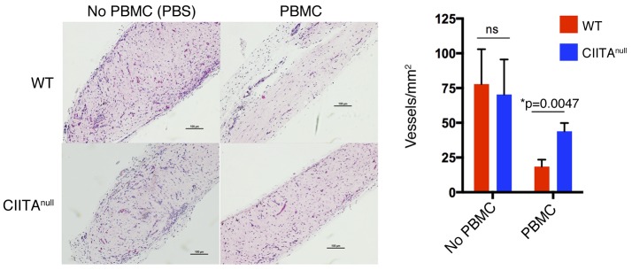
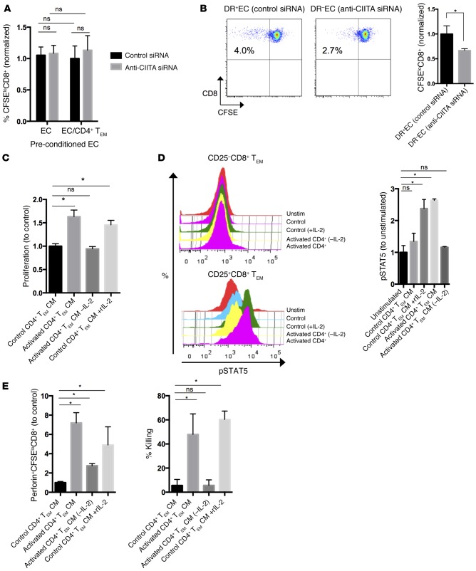
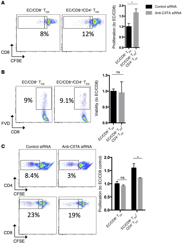
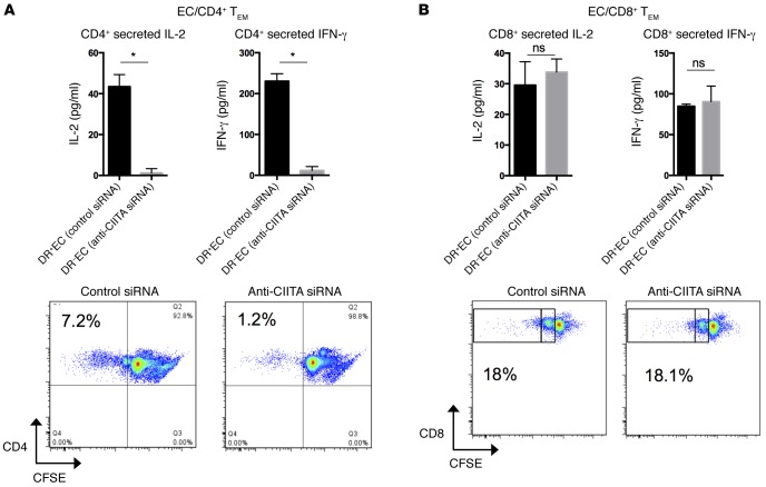
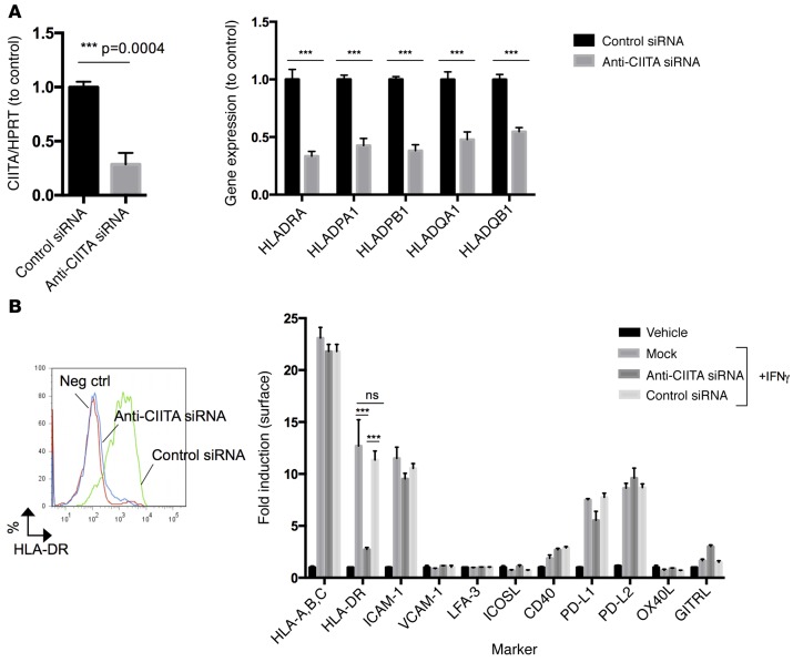
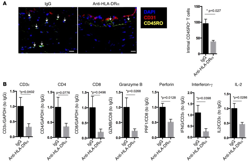
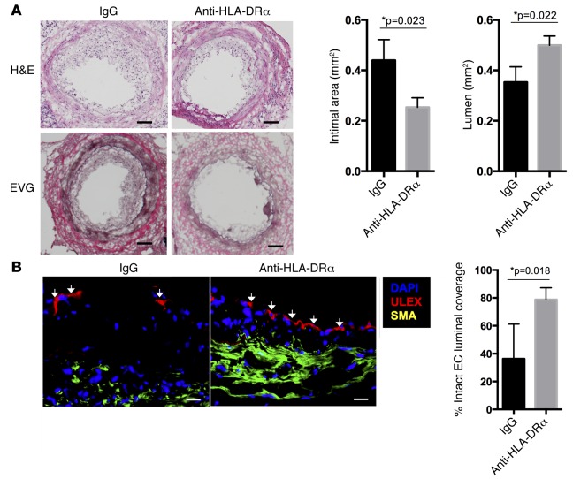
Acute allograft rejection is mediated by host CD8+ cytotoxic T lymphocytes (CTL) targeting graft class I major histocompatibility complex (MHC) molecules. In experimental rodent models, rejection requires differentiation of naive CD8+ T cells into alloreactive CTL within secondary lymphoid organs, whereas in humans, CTL may alternatively develop within the graft from circulating CD8+ effector memory T cells (TEM) that recognize class I MHC molecules on graft endothelial cells (EC). This latter pathway is poorly understood. Here, we show that host CD4+ TEM, activated by EC class II MHC molecules, provide critical help for this process. First, blocking HLA-DR on EC lining human artery grafts in immunodeficient mice reduces CD8+ CTL development within and acute rejection of the artery by adoptively transferred allogeneic human lymphocytes. Second, siRNA knockdown or CRISPR/Cas9 ablation of class II MHC molecules on EC prevents CD4+ TEM from helping CD8+ TEM to develop into CTL in vitro. Finally, implanted synthetic microvessels, formed from CRISPR/Cas9-modified EC lacking class II MHC molecules, are significantly protected from CD8+ T cell-mediated destruction in vivo. We conclude that human CD8+ TEM-mediated rejection targeting graft EC class I MHC molecules requires help from CD4+ TEM cells activated by recognition of class II MHC molecules.


Figures

Figure 7
Figure 7. Genetic ablation of CIITA in EC by CRISPR/Cas9 reduces T cell–mediated destruction of synthetic vessels in vivo.
Synthetic human microvessels formed from WT or CIITAnull EC were implanted s.c. in the abdominal wall of C.B-17 SCID/bg mice. After 2 weeks, mice were adoptively transferred with peripheral blood mononuclear cells (PBMC). In the absence of circulating T cells, WT and CIITAnull EC form equivalent numbers of vessels (n = 3 each). In the presence of circulating T cells, however, there is a significantly higher loss of perfused vessels formed from WT compared with CIITAnull EC (n = 3 each). Scale bar: 100 μm. *P < 0.05, 2-tailed Student’s t test. CIITA, class II MHC transactivator; bg, beige; EC,endothelial cells.
Figure 6
Figure 6. CD4+ TEM enhancement of CD8+ TEM responses are mediated by secreted IL-2.
(A) EC precultured with CD4+ TEM and subsequently isolated by FACS have not increased their capacity to activate CD8+ TEM. (B) When separated by a semipermeable transwell membrane, CD4+ TEM enhance CD8+ TEM alloresponses. (C) CM generated by EC-activated CD4+ TEM is sufficient to enhance proliferation of CD8+ TEM. Immunoabsorption of IL-2 removes that capacity, whereas addition of IL-2 to control CM mimics activated CM. (D) Similarly, CM induces phosphorylates STAT5 in CD25+ but not CD25CD8+ TEM in an IL-2–dependent manner. (E) In addition, CM enhances expression of perforin and promotes CTL killing in an IL-2–dependent manner. Similar results were seen in three independent experiments. *P < 0.05, by 2-tailed Student’s t test (B) or by 1-way ANOVA with Bonferroni post-hoc test (A and CE). TEM, effector memory T cell; CIITA, class II MHC transactivator; CM, conditioned medium; EC,endothelial cells; CTL, cytotoxic T cell.
Figure 5
Figure 5. Activation of CD4+ TEM by allogeneic EC is necessary to enhance CD8+ TEM responses to the same EC.
(A and B) Addition of CD4+ TEM to EC/CD8+ TEM cocultures enhances CD8+ TEM expansion as measured by CFSE dilution and flow cytometry at 7 days (A), but CD4+ TEM do not enhance survival of CD8+ TEM (B) as measured by viability. (C) Knockdown of CIITA expression by siRNA, to prevent CD4+ TEM activation, inhibits CD4+ TEM enhancement of CD8+ TEM expansion at 7 days as measured by CFSE dilution and flow cytometric analysis at 7 days. Note that the inclusion of activated CD4+ TEM increases the rounds of replication of CD8+ TEM (inset boxes). Similar results were seen in three independent experiments. *P < 0.05, 2-tailed Student’s t test. TEM, effector memory T cell; CIITA, class II MHC transactivator; FVD, fixable viability dye; EC,endothelial cells.
Figure 4
Figure 4. siRNA-mediated knockdown of CIITA in EC directly inhibits CD4+ but not CD8+ TEM alloresponses.
(A and B) Anti-CIITA siRNA inhibits activation of CD4+ TEM (10:1 T cell/EC ratio) (A) but not activation of CD8+ TEM (30:1 T cell/EC ratio) (B), as measured by ELISA at 24 hours for IL-2 and IFN-γ and proliferation by CFSE dilution and flow cytometric analysis at 7 days. Note that the majority of CD8+ TEM have divided only once, whereas CD4+ TEM undergo multiple rounds of proliferation. Similar results were seen in three independent experiments. *P < 0.05, 2-tailed Student’s t test. CIITA, class II MHC transactivator; TEM, effector memory T cell; EC, endothelial cells.
Figure 3
Figure 3. anti-CIITA siRNA selectively inhibits induction of CIITA and class II MHC molecules in human EC.
Cultured human EC were transfected with anti-CIITA siRNA or control siRNA on day 0, treated with IFN-γ at 24 hours, and analyzed on day 3. (A and B) Specific transcripts were measured by qPCR (A), and protein was assessed by FACS (B). Similar results were seen in four independent experiments. ***P < 0.0005, 2-tailed Student’s t test. CIITA, class II MHC transactivator; MHC, major histocompatibility complex; EC, endothelial cells.
Figure 2
Figure 2. HLA-DR blockade reduces CD8+ T cell infiltration into and CTL development within implanted allogeneic vessel segments in vivo.
(A and B) Anti–HLA-DRα F(ab)′2 fragment significantly (n = 6 per group) reduces total intimal T cell infiltration as detected by immunofluorescence (scale bar: 20 μm) (A) and CD8+ T cell–associated transcripts of cytokines and CTL effector molecules in rejected artery grafts at 21 days (B) as detected by qPCR normalized to either total mRNA or T cell–specific mRNA as indicated on the y axis. Arrows indicate infiltrating intimal CD45RO+ T cells. *P < 0.05, 2-tailed Student’s t test. CTL, cytotoxic T cell.
Figure 1
Figure 1. HLA-DR blockade reduces acute T cell–mediated injury to implanted allogeneic vessel segments in vivo.
(A and B) In an MHC-mismatched model of arterial rejection by allogeneic T cells (n = 6 per group), blockade of class II MHC by anti–HLA-DRα F(ab)′2 fragment reduces intimal area expansion and increases lumen area as measured by H&E and EVG staining (scale bar: 50 μm) (A) and reduces disruption of EC lining the vessel lumen, a hallmark of intimal arteritis/endothelialitis, as measured by percent of circumferential coverage (scale bar: 20 μm) (B), both at 21 days. Arrows indicate areas of intact endothelial lining. *P < 0.05, 2-tailed Student’s t test. EVG, Elastia-van Gieson; MHC, major histocompatibility complex; EC, endothelial cells.

References

    1. Suthanthiran M, Strom TB. Renal transplantation. N Engl J Med. 1994;331(6):365–376. doi: 10.1056/NEJM199408113310606. - DOI - PubMed
    1. Stehlik J, et al. The Registry of the International Society for Heart and Lung Transplantation: Twenty-eighth Adult Heart Transplant Report — 2011. J Heart Lung Transplant. 2011;30(10):1078–1094. doi: 10.1016/j.healun.2011.08.003. - DOI - PubMed
    1. Taylor DO, et al. Registry of the International Society for Heart and Lung Transplantation: Twenty-sixth Official Adult Heart Transplant Report — 2009. J Heart Lung Transplant. 2009;28(10):1007–1022. doi: 10.1016/j.healun.2009.08.014. - DOI - PubMed
    1. Colvin RB, et al. Evaluation of pathologic criteria for acute renal allograft rejection: reproducibility, sensitivity, and clinical correlation. J Am Soc Nephrol. 1997;8(12):1930–1941. - PubMed
    1. Strehlau J, et al. Quantitative detection of immune activation transcripts as a diagnostic tool in kidney transplantation. Proc Natl Acad Sci U S A. 1997;94(2):695–700. doi: 10.1073/pnas.94.2.695. - DOI - PMC - PubMed