

Brain Atrophy Correlates with Severe Enlarged Perivascular Spaces in Basal Ganglia among Lacunar Stroke Patients
- PMID: 26900696
- PMCID: PMC4764761
- DOI: 10.1371/journal.pone.0149593
Brain Atrophy Correlates with Severe Enlarged Perivascular Spaces in Basal Ganglia among Lacunar Stroke Patients
Abstract
Background: Enlarged perivascular spaces (EPVS) correlate with cognitive impairment and incident dementia. However, etiologies for severe basal ganglia EPVS (BG-EPVS) are still unclear. Our aim was to investigate the independent risk factors for severe BG-EPVS in patients with acute lacunar stroke.
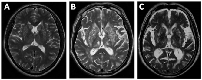
Methods: We prospectively identified patients with lacunar stroke (diameter on DWI ≤ 20mm) from Jan 2011 to May 2015. Patients with severe BG-EPVS were identified on T2 weighted MRI. Age (± 1 year) and sex matched controls were also recruited in the same population (two controls for one case). Vascular risk factors, clinical data, EPVS in centrum semiovale (rated 0 to 4), white matter hyperintensities (WMH) (by Fazekas scale), brain atrophy (rated 0 to 6) were compared between two groups. Logistic regression was performed to determine independent risk factors for severe BG-EPVS.
Results: During study period, 89 patients with severe BG-EPVS and 178 matched controls were included. Vascular risk factors did not differ between two groups. Patients with severe BG-EPVS had lower level of HbA1c and diastolic BP at admission, but presented with larger infarct size, more severe WMH (including total WMH, periventricular WMH and deep WMH) and brain atrophy. In logistic regression, brain atrophy (OR = 1.40; 95%CI 1.13, 1.73) and deep WMH (OR = 1.88; 95%CI 1.24, 2.83) were independent risk factors for severe BG-EPVS.
Conclusions: Brain atrophy and deep WMH are independent risk factors for severe BG-EPVS, supporting the hypothesis that brain atrophy may be associated with the development of EPVS in basal ganglia.
Conflict of interest statement
Figures
References
-
- Kwee RM, Kwee TC. Virchow-robin spaces at mr imaging. Radiographics 2007; 27: 1071–1086. - PubMed
Publication types
MeSH terms
LinkOut - more resources
Full Text Sources
Other Literature Sources
