Review
. 2016 May;54(5):636-46.
doi: 10.1165/rcmb.2016-0045PS.

Autophagy and Obesity-Related Lung Disease

Affiliations
Review

Autophagy and Obesity-Related Lung Disease

Maria A Pabon et al. Am J Respir Cell Mol Biol. 2016 May.

Abstract

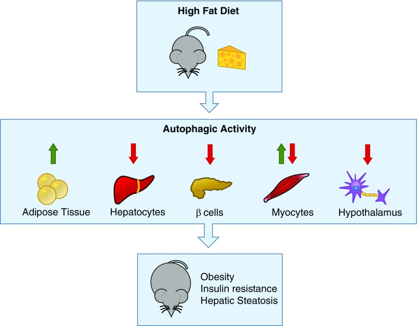
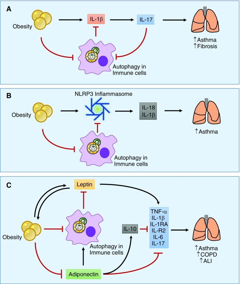
Obesity-related disease is a significant source of premature death and economic burden globally. It is also a common comorbidity in patients suffering from lung disease, affecting both severity and treatment success. However, this complex association between obesity and the lung is poorly understood. Autophagy is a self-recycling homeostatic process that has been linked to beneficial or deleterious effects, depending on the specific lung disease. Obesity affects autophagy in a tissue-specific manner, activating autophagy in adipocytes and impairing autophagy in hepatocytes, immune cells, and pancreatic β-cells, among others. Obesity is also characterized by chronic low-grade inflammation that can be modulated by the pro- and antiinflammatory effects of the autophagic machinery. Scant evidence exists regarding the impact of autophagy in obesity-related lung diseases, but there are communal pathways that could be related to disease pathogenesis. Important signaling molecules in obesity, including IL-17, leptin, adiponectin, NLRP3 inflammasome, and TLR-4, have been implicated in the pathogenesis of lung disease. These mediators are known to be modulated by autophagy activity. In this perspective, we highlight the recent advances in the understanding of autophagy in obesity-related conditions, as well as the potential mechanisms that can link autophagy and obesity in the pathogenesis of lung disease.

Keywords: autophagy; inflammation; lipid metabolism; lung disease; obesity.


Figures

Figure 1.
Figure 1.
Molecular mechanism of autophagy. Environmental signals modulate mammalian target of rapamycin (mTOR) complex 1 (mTORC1), negatively regulating autophagy by inhibiting the uncoordinated-51–like kinase 1 (ULK1) complex consisting of ULK1, ATG101, ATG13, and FIP200. Starvation and low ATP levels down-regulate mTOR and directly stimulate the ULK1 complex. The ULK1 complex positively regulates autophagy by activating the Beclin 1 interacting complex, which consists of Beclin 1 (BCL2 family proteins), VPS34 (a class III phosphatidylinositol-3 kinase), and ATG14L. This increases the levels of phosphatidylinositol 3-phosphate (PI3P), which promotes the nucleation of autophagosomal membrane. The elongation of the autophagosome membrane requires two ubiquitin-like conjugation systems. The first is the ATG5–ATG12 complex, which is conjugated by ATG7 and ATG10 enzymes. The second one requires the ubiquitin-like protein microtubule–associated protein 1 light chain 3 (LC3), also called ATG8, which is cleaved by ATG4B into LC3B-I. LC3B-I turns into the active LC3B-II after conjugation with phosphatidylethanolamine by ATG3 and ATG7. Once the double-membrane autophagosome is complete, it fuses with a lysosome to form the autophagolysosome to degrade the autophagosome contents. ATG, autophagy-related protein; FIP200, focal adhesion kinase family interacting protein of 200 kD; VPS34, vacuolar protein sorting 34.
Figure 2.
Figure 2.
Role of autophagy in lipid metabolism in the liver. In hepatocytes, autophagy plays an important role in lipid turnover from lipid droplets. In starvation, autophagy degrades lipid droplets to increase free fatty acids and fuel β-oxidation. In obesity-related conditions such as hyperinsulinemia and lipid accumulation, autophagy is inhibited, which causes a predisposition toward more lipid accumulation and, in turn, further autophagy inhibition that, in organs such as the liver, can lead to hepatic steatosis.
Figure 3.
Figure 3.
Tissue-specific regulation of autophagy under high-fat diet conditions. Under high-fat diet conditions, mice have tissue-specific changes in autophagy. In adipose tissue, there is an increase in autophagic activity as a response to endoplasmic reticulum stress, leading to degradation of the antiinflammatory adipokine adiponectin. In hepatocytes, β cells, and hypothalamic neurons, there is decreased autophagy under a high-fat diet, leading to lipid accumulation, β-cell toxicity, and inflammation. In myocytes under exercise, there is an increase in autophagy, leading to decreased insulin resistance. Thus, aberrant autophagy contributes to obesity disease pathogenesis, leading to insulin resistance, hepatic steatosis, and inflammation.
Figure 4.
Figure 4.
Proposed mechanisms of obesity- and autophagy-related pathogenesis of lung disease. (A) Obesity induces the production of inflammatory cytokines such as IL-1β by macrophages in the adipose or lung tissue, leading to the production of IL-17. IL-17 has been correlated with worsening lung inflammation and injury in diseases such as asthma and fibrosis. Autophagy can sequester pro–IL-1β, decreasing IL-1β production and thus negatively regulating IL-17 levels. (B) Obesity is characterized by NLRP3 inflammasome activation that increases the production of inflammatory cytokines such as IL-1β and IL-18, which has been shown to contribute to lung disease pathogenesis. Autophagy can inhibit inflammasome activation, thereby decreasing IL-1β and IL-18 production. (C) Adipocytes are characterized by adipokine production such as leptin and adiponectin. Under obesity conditions, leptin levels are increased as a result of leptin resistance. Leptin has systemic effects and can increase the production of inflammatory cytokines. Adiponectin is decreased in obesity. Adiponectin has antiinflammatory properties through increasing production of IL-10 and inhibits the production of proinflammatory cytokines such as TNF-α, IL-1β, IL-1RA, IL-R2, IL-6, and IL-17. ALI, acute lung injury; COPD, chronic obstructive pulmonary disease; NLRP3, nod-like receptor protein-3.

References

    1. World Health OrganizationObesity and overweight [Internet]. World Health Organization; 2015. [accessed 10 Jan 2016]Available from: http://www.who.int/mediacentre/factsheets/fs311/en/
    1. McNeil H, Segal L Quality of Life and Obesity. The Centre for Health Program Evaluation Monash University. 1999. Research report 17.
    1. Strong AL, Burow ME, Gimble JM, Bunnell BA. Concise review: the obesity cancer paradigm: exploration of the interactions and crosstalk with adipose stem cells. Stem Cells. 2015;33:318–326. - PMC - PubMed
    1. Versini M, Jeandel PY, Rosenthal E, Shoenfeld Y. Obesity in autoimmune diseases: not a passive bystander. Autoimmun Rev. 2014;13:981–1000. - PubMed
    1. Karrowni W, Kennedy K, Jones P. Obesity paradox among survivors of acute myocardial infarction and its interaction with time. J Am Coll Cardiol. 2015;65(10S):A31.