. 2016 Feb 22;11(2):e0147358.
doi: 10.1371/journal.pone.0147358. eCollection 2016.

Specific Neuropilins Expression in Alveolar Macrophages among Tissue-Specific Macrophages

Affiliations

Specific Neuropilins Expression in Alveolar Macrophages among Tissue-Specific Macrophages

Naing Ye Aung et al. PLoS One. .

Abstract

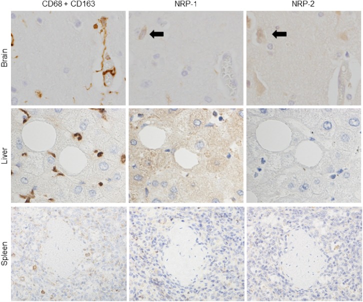
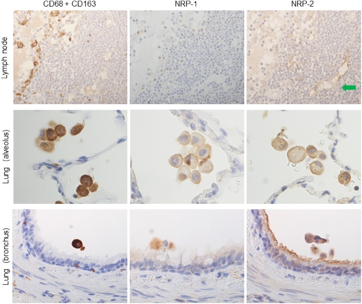
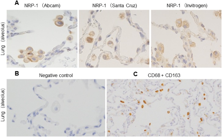
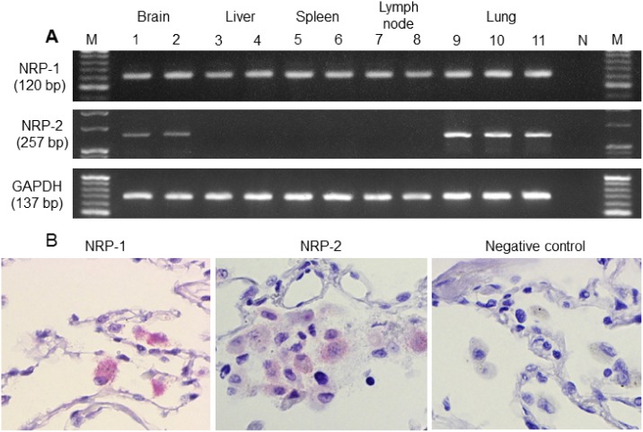
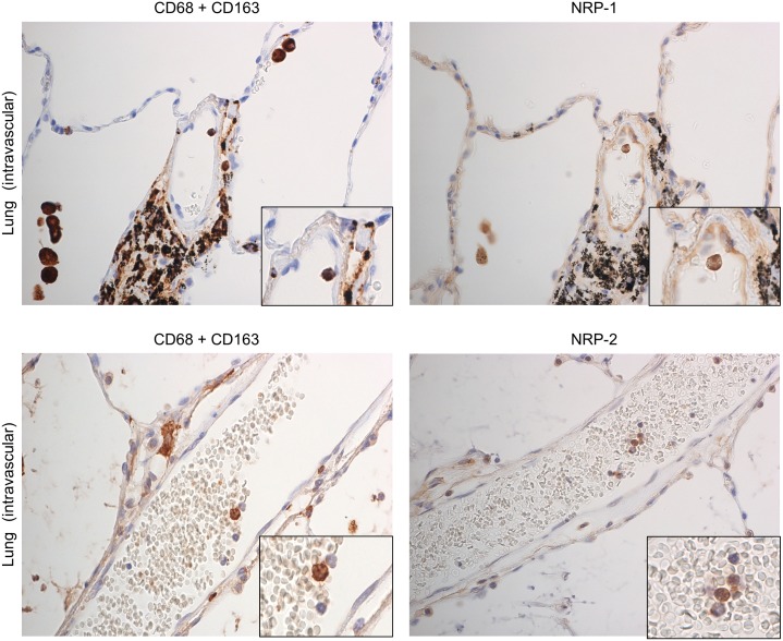
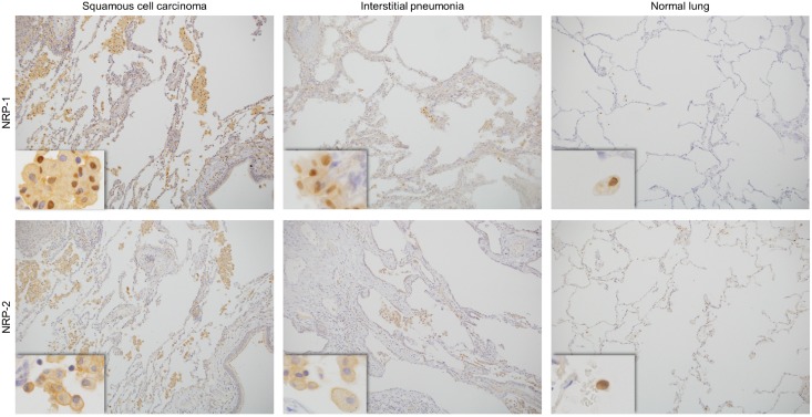
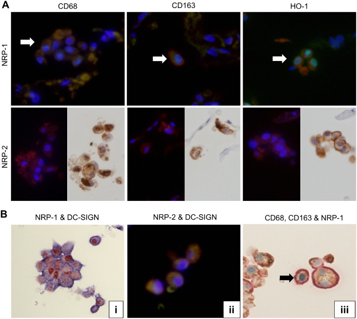
In the immune system, neuropilins (NRPs), including NRP-1 and NRP-2, are expressed in thymocytes, dendritic cells, regulatory T cells and macrophages. Their functions on immune cells around the neoplastic cells vary into pro-angiogenesis, tumor progression and anti-angiogenesis according to their ligands. Even though NRPs expression on malignant tumors and immune system has studied, a PubMed-based literature query did not yield any articles describing NRPs expression on tissue-specific macrophages. The aims of this study were (i) to detect NRPs expression on tissue-specific macrophages in the brain, liver, spleen, lymph node and lung; (ii) to observe NRPs expression in classes of macrophages, including alveolar macrophages (AMs), bronchial macrophages (BMs), interstitial macrophages (IMs), intravascular macrophages (IVMs) and macrophage subsets (M1, M2 and Mox) in lung; and (iii) to detect the co-expression of NRPs and dendritic cell-specific ICAM-3-grabbing nonintegrin (DC-SIGN) in AMs. Both NRPs were specifically detected in AMs among tissue-specific macrophages by immunohistochemistry (IHC). NRPs mRNA expression levels were characterized in normal lung by reverse transcriptase polymerase chain reaction (RT-PCR) and in situ-polymerase chain reaction (in situ-PCR). The expression of both NRPs was detected in AMs, BMs and IVMs by IHC. The frequency of NRPs+ AMs in lung tissue adjacent to the cancer margin was significantly higher than the frequencies in inflamed and normal lung tissue. Double and triple IHC demonstrated that NRPs are expressed on all macrophage subsets in lung. Double IHC showed co-expression of DC-SIGN and NRPs in AMs. This study demonstrated for the first time the specific expression of both NRPs in AMs among tissue-specific macrophages and their expression on M1, M2 and Mox macrophages. Furthermore, the possible origin of AMs from blood monocytes could be suggested from a co-expression of NRPs and DC-SIGN.


Conflict of interest statement

Competing Interests: The authors have declared that no competing interests exist.

Figures

Fig 1
Fig 1. NRPs expression in tissue-specific macrophages compared to immunostaining with a cocktail of anti-CD68 and anti-CD163 antibodies.
Tissue-specific macrophages were recognized by immunostaining with a cocktail of anti-CD68 and CD163 antibodies in serial sections. NRP-1 and NRP-2 expression was not observed in tissue-specific macrophages of brain (microglia), liver (Kupffer cells) and spleen (red pulp macrophages). Black arrows indicate the neuronal staining of NRPs in brain. Serial sections were counterstained with hematoxylin. NRP-1, neuropilin 1; NRP-2, neuropilin 2.
Fig 2
Fig 2. NRPs expression in tissue-specific macrophages compared to immunostaining with a cocktail of anti-CD68 and anti-CD163 antibodies.
Tissue-specific macrophages were recognized by immunostaining with a cocktail of anti-CD68 and CD163 antibodies in serial sections. NRP-1 and NRP-2 expression was detected in alveolar macrophages in lung, but not in lymph node (sinus macrophages). And NRP-1 and NRP-2 also expressed on bronchial macrophages. Green arrow indicates NRP-2 expression on lymphatic vascular endothelium, used as positive control. Serial sections were counterstained with hematoxylin. NRP-1, neuropilin 1; NRP-2, neuropilin 2.
Fig 3
Fig 3. NRPs expression from different sources in alveolar macrophages and, IHC with a cocktail of anti-CD68 and anti-CD163 antibodies in serial section of in-situ PCR.
(A) Immunohistochemistry showed alveolar macrophages expressing NRP-1 as detected by using 3 different antibodies from Abcam, Santa Cruz Biotechnology and Invitrogen. Serial sections were counterstained with hematoxylin. (B) Isotype or negative control showed no reactivity. It was counterstained with hematoxylin. (C) All alveolar macrophages showed positivity with CD68 and CD163. NRP-1, neuropilin 1.
Fig 4
Fig 4. NRPs mRNAs expression in normal tissues (RT-PCR) and on alveolar macrophages in physiologically normal lung (in situ-PCR).
(A) By reverse transcriptase polymerase chain reaction (RT-PCR), NRP-1 mRNA was expressed in normal brain (lanes 1, 2), liver (lanes 3, 4), spleen (lanes 5, 6), lymph node (lanes 7, 8) and lung (lanes 9, 10 and 11). NRP-2 mRNA was expressed in normal lung and brain but was not expressed in liver, spleen and lymph node. N represents the negative control, and M represents the 20 base-pair DNA ladder. (B) NRP-1 and NRP-2 mRNAs of alveolar macrophages in physiologically normal lung (remote to the cancer nest), as observed by in situ-polymerase chain reaction (in situ-PCR). NRP-1, neuropilin 1; NRP-2, neuropilin 2; GAPDH, glyceraldehyde-3-phosphate dehydrogenase.
Fig 5
Fig 5. NRPs expression in intravascular macrophages in physiologically normal lung by immunostaining.
Intravascular macrophages (blood monocytes) were recognized by immunostaining with a cocktail of anti-CD68 and CD163 antibodies. NRP-1 and NRP-2 expression was observed in intravascular macrophages. Green arrow indicates NRP-1 expression on vascular endothelium, used as positive control. Serial sections were counterstained with hematoxylin. NRP-1, neuropilin 1; NRP-2, neuropilin 2.
Fig 6
Fig 6. Immunohistochemistry of NRPs expression on alveolar macrophages in lung tissue (adjacent to the cancer margin, inflamed and physiologically normal).
NRP-1 and NRP-2 expressed on alveolar macrophages in lung tissue adjacent to cancer (squamous cell carcinoma), inflamed lung (interstitial pneumonia) and physiologically normal lung (remote to the cancer nest). NRP-1, neuropilin 1; NRP-2, neuropilin 2.
Fig 7
Fig 7. Double immunostaining (NRPs vs CD68, CD163, HO-1 and DC-SIGN) and triple immunohistochemistry (CD68, CD163 and NRP-1) of lung tissue adjacent to the cancer.
(A) Double IF showed expression of CD68, CD163 and HO-1 (Rhodamine, anti-mouse, red color) on NRP-1+(Fluorescein, anti-rabbit, green color) alveolar macrophages. White arrows showed double positive cells. Single IHC after single IF showed NRP-2+ (Rhodamine, anti-mouse, red color) alveolar macrophages also express CD68, CD163 and HO-1 (LSAB, anti-mouse, brown color). (B) (i) Double IHC showed NRP-1(Red) and DC-SIGN (blue) positive alveolar macrophages. (ii) Double IF showed the co-expression of NRP-2 (Rhodamine, anti-mouse, red color) and DC-SIGN (Fluorescein, anti-rabbit, green color) on AMs. (iii) Triple IHC of CD68 (brown), CD163 (light red) and NRP-1 (light blue) showed triple-positive cells (CD68+CD163+NRP-1+; indicated by black arrow) and double-positive cells (CD68+NRP-1+, CD68+CD163+ and CD163+NRP-1+). NRP-1, neuropilin 1; NRP-2, neuropilin 2; DC-SIGN, dendritic cell-specific ICAM-3-grabbing nonintegrin.

References

    1. Fujisawa H. Discovery of semaphorine receptors, neuropilin and plexin, and their functions in neural development. J Neurobiol. 2004;59:24–33. - PubMed
    1. Pellet-Many C, Frankel P, Jia H, Zachary I.Neuropilins: structure, function and role in disease. Biochem J. 2008;411:211–226. 10.1042/BJ20071639 - DOI - PubMed
    1. Raimondi C, Ruhrberg C. Neuropilin signalling in vessels, neurons and tumours. Semin Cell Dev Biol. 2013;24:172–178. 10.1016/j.semcdb.2013.01.001 - DOI - PubMed
    1. West DC, Rees CG, Duchesne L, Patey SJ, Terry CJ, Turnbull JE, et al. Interactions of multiple heparin binding growth factors with neuropilin-1 and potentiation of the activity of fibroblast growth factor-2. J Biol Chem. 2005;280:13457–13464. - PubMed
    1. Hsieh SH, Ying NW, Wu MH, Chiang WF, Hsu CL, Wong TY, et al. Galectin-1, a novel ligand of neuropilin-1, activates VEGFR-2 signaling and modulates the migration of vascular endothelial cells. Oncogene. 2008;27:3746–3753. 10.1038/sj.onc.1211029 - DOI - PubMed

Publication types