

In Vivo Screening Using Transgenic Zebrafish Embryos Reveals New Effects of HDAC Inhibitors Trichostatin A and Valproic Acid on Organogenesis
- PMID: 26900852
- PMCID: PMC4763017
- DOI: 10.1371/journal.pone.0149497
In Vivo Screening Using Transgenic Zebrafish Embryos Reveals New Effects of HDAC Inhibitors Trichostatin A and Valproic Acid on Organogenesis
Abstract
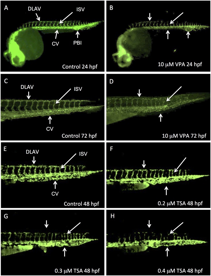
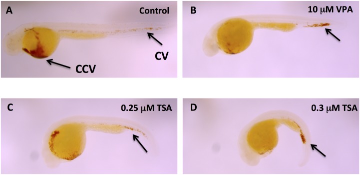
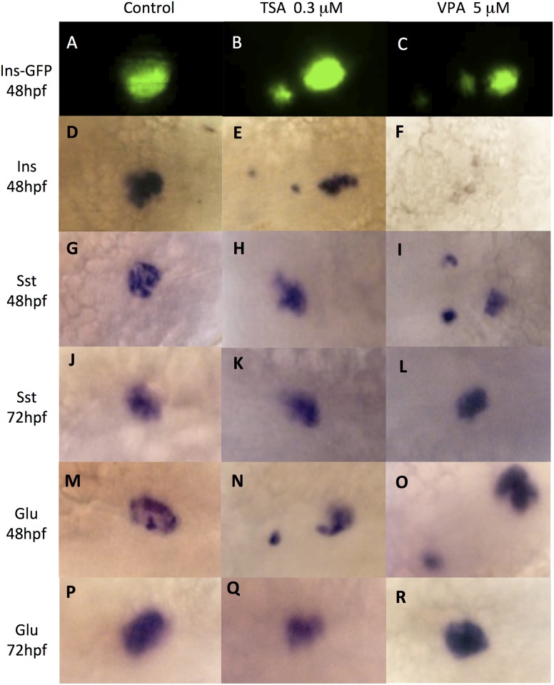
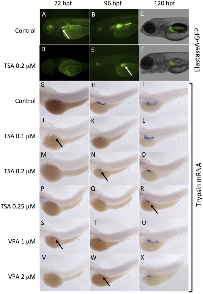
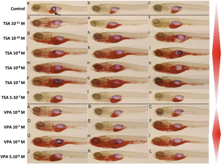
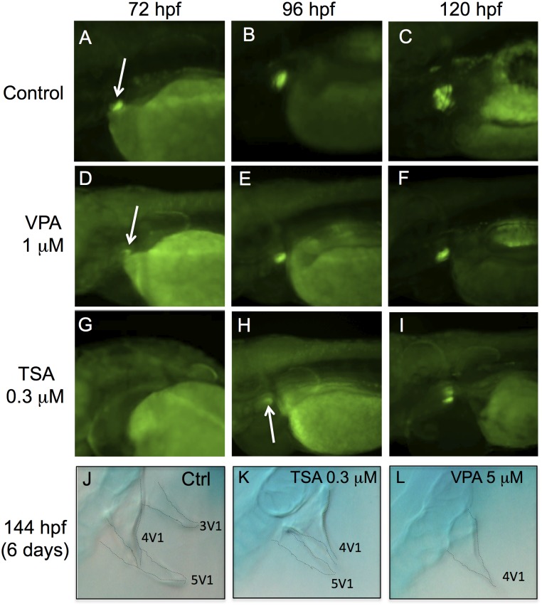
The effects of endocrine disrupting chemicals (EDCs) on reproduction are well known, whereas their developmental effects are much less characterized. However, exposure to endocrine disruptors during organogenesis may lead to deleterious and permanent problems later in life. Zebrafish (Danio rerio) transgenic lines expressing the green fluorescent protein (GFP) in specific organs and tissues are powerful tools to uncover developmental defects elicited by EDCs. Here, we used seven transgenic lines to visualize in vivo whether a series of EDCs and other pharmaceutical compounds can alter organogenesis in zebrafish. We used transgenic lines expressing GFP in pancreas, liver, blood vessels, inner ear, nervous system, pharyngeal tooth and pectoral fins. This screen revealed that four of the tested chemicals have detectable effects on different organs, which shows that the range of effects elicited by EDCs is wider than anticipated. The endocrine disruptor tetrabromobisphenol-A (TBBPA), as well as the three drugs diclofenac, trichostatin A (TSA) and valproic acid (VPA) induced abnormalities in the embryonic vascular system of zebrafish. Moreover, TSA and VPA induced specific alterations during the development of pancreas, an observation that was confirmed by in situ hybridization with specific markers. Developmental delays were also induced by TSA and VPA in the liver and in pharyngeal teeth, resulting in smaller organ size. Our results show that EDCs can induce a large range of developmental alterations during embryogenesis of zebrafish and establish GFP transgenic lines as powerful tools to screen for EDCs effects in vivo.
Conflict of interest statement
Figures
Similar articles
-
Mta3-NuRD complex is a master regulator for initiation of primitive hematopoiesis in vertebrate embryos.Blood. 2009 Dec 24;114(27):5464-72. doi: 10.1182/blood-2009-06-227777. Epub 2009 Oct 28. Blood. 2009. PMID: 19864643
-
Association of valproate-induced teratogenesis with histone deacetylase inhibition in vivo.FASEB J. 2005 Jul;19(9):1166-8. doi: 10.1096/fj.04-3425fje. Epub 2005 May 18. FASEB J. 2005. PMID: 15901671
-
Perfluorobutanesulfonic Acid Disrupts Pancreatic Organogenesis and Regulation of Lipid Metabolism in the Zebrafish, Danio rerio.Toxicol Sci. 2019 Jan 1;167(1):258-268. doi: 10.1093/toxsci/kfy237. Toxicol Sci. 2019. PMID: 30239974 Free PMC article.
-
Gal4 Driver Transgenic Zebrafish: Powerful Tools to Study Developmental Biology, Organogenesis, and Neuroscience.Adv Genet. 2016;95:65-87. doi: 10.1016/bs.adgen.2016.04.002. Epub 2016 Jun 13. Adv Genet. 2016. PMID: 27503354 Review.
-
Impact of endocrine-disrupting chemicals on reproductive function in zebrafish (Danio rerio).Reprod Domest Anim. 2015 Feb;50(1):1-6. doi: 10.1111/rda.12468. Epub 2014 Dec 22. Reprod Domest Anim. 2015. PMID: 25529055 Review.
Cited by
-
The transcription factor, Nuclear factor, erythroid 2 (Nfe2), is a regulator of the oxidative stress response during Danio rerio development.Aquat Toxicol. 2016 Nov;180:141-154. doi: 10.1016/j.aquatox.2016.09.019. Epub 2016 Oct 1. Aquat Toxicol. 2016. PMID: 27716579 Free PMC article.
-
Potential of the zebrafish (Danio rerio) embryo test to discriminate between chemicals of similar molecular structure-a study with valproic acid and 14 of its analogues.Arch Toxicol. 2022 Nov;96(11):3033-3051. doi: 10.1007/s00204-022-03340-z. Epub 2022 Aug 3. Arch Toxicol. 2022. PMID: 35920856 Free PMC article.
-
Comparative study of herbal plants on the phenolic and flavonoid content, antioxidant activities and toxicity on cells and zebrafish embryo.J Tradit Complement Med. 2017 Jan 16;7(4):452-465. doi: 10.1016/j.jtcme.2016.12.006. eCollection 2017 Oct. J Tradit Complement Med. 2017. PMID: 29034193 Free PMC article.
-
Histone deacetylase activity mediates thermal plasticity in zebrafish (Danio rerio).Sci Rep. 2019 Jun 3;9(1):8216. doi: 10.1038/s41598-019-44726-x. Sci Rep. 2019. PMID: 31160672 Free PMC article.
-
Comparative toxicological assessment of 2 bisphenols using a systems approach: evaluation of the behavioral and transcriptomic responses of Danio rerio to bisphenol A and tetrabromobisphenol A.Toxicol Sci. 2024 Aug 1;200(2):394-403. doi: 10.1093/toxsci/kfae063. Toxicol Sci. 2024. PMID: 38730555 Free PMC article.
References
-
- Carson RL (1962) Silent spring: Houghton Mifflin.
-
- Colborn T, Dumanoski D, Myers JP (1996) Our stolen future: Dutton.
Publication types
MeSH terms
Substances
LinkOut - more resources
Full Text Sources
Other Literature Sources
Molecular Biology Databases
Research Materials
