. 2016 Apr;12(4):282-9.
doi: 10.1038/nchembio.2036. Epub 2016 Feb 22.

Autopalmitoylation of TEAD proteins regulates transcriptional output of the Hippo pathway

Affiliations

Autopalmitoylation of TEAD proteins regulates transcriptional output of the Hippo pathway

PuiYee Chan et al. Nat Chem Biol. 2016 Apr.

Abstract

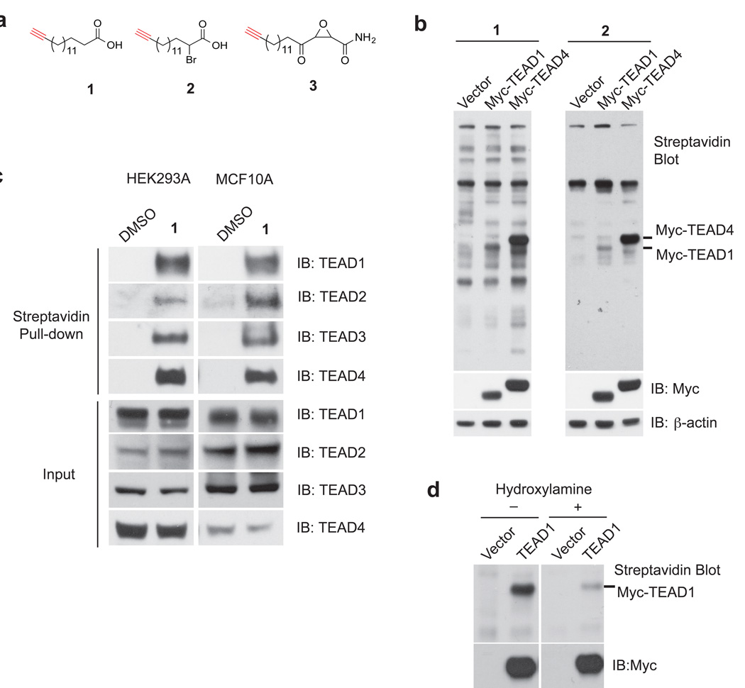
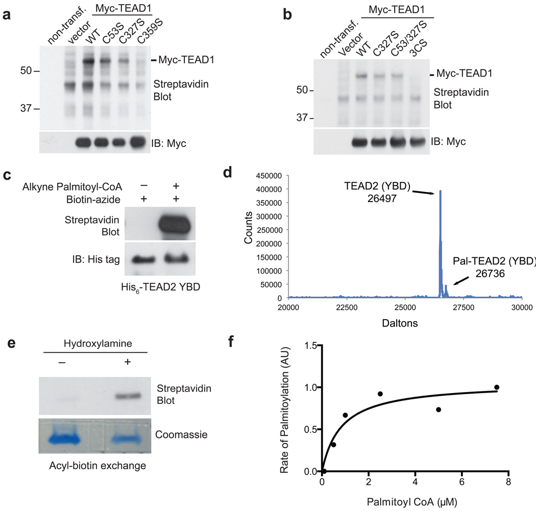
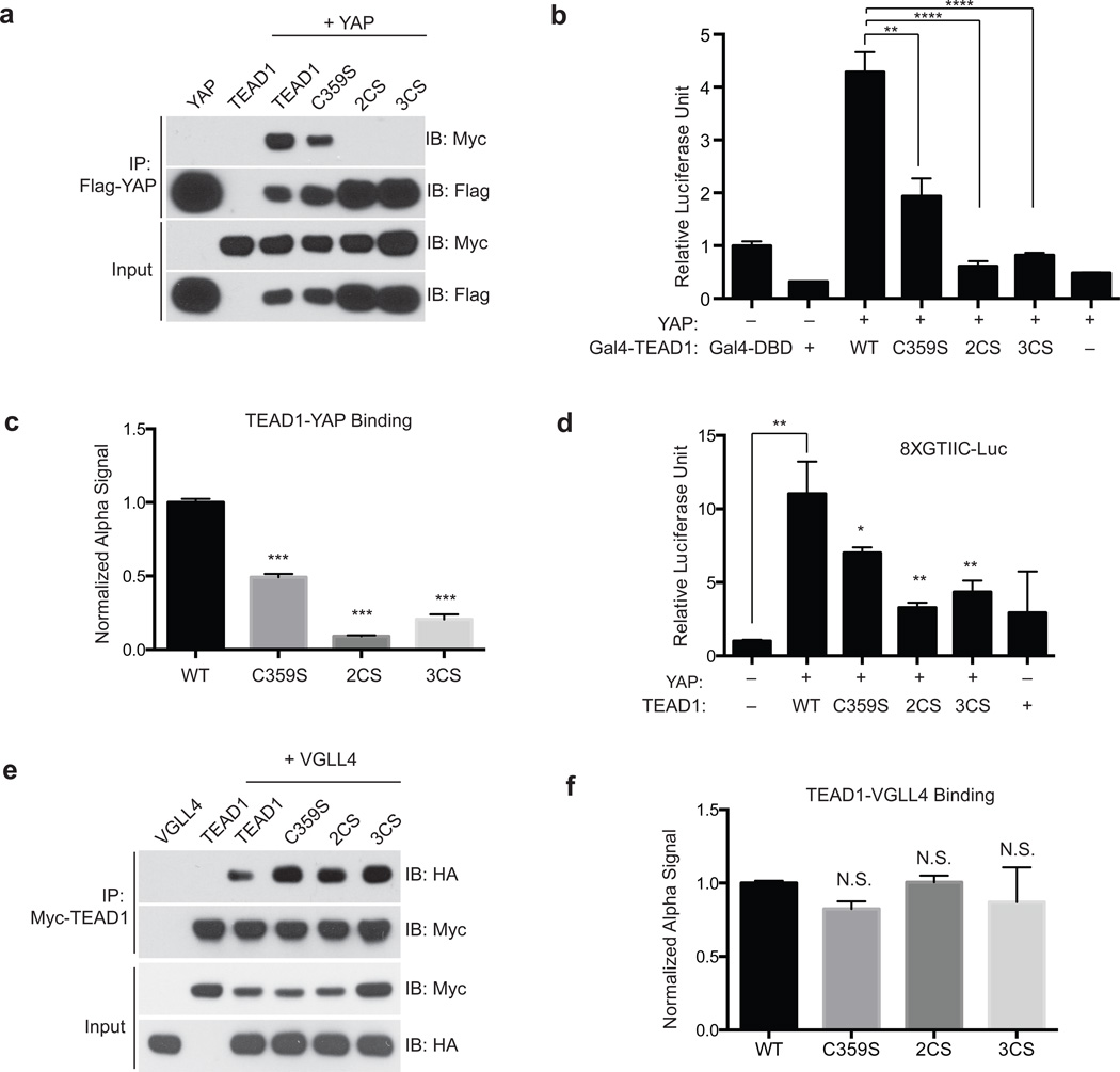
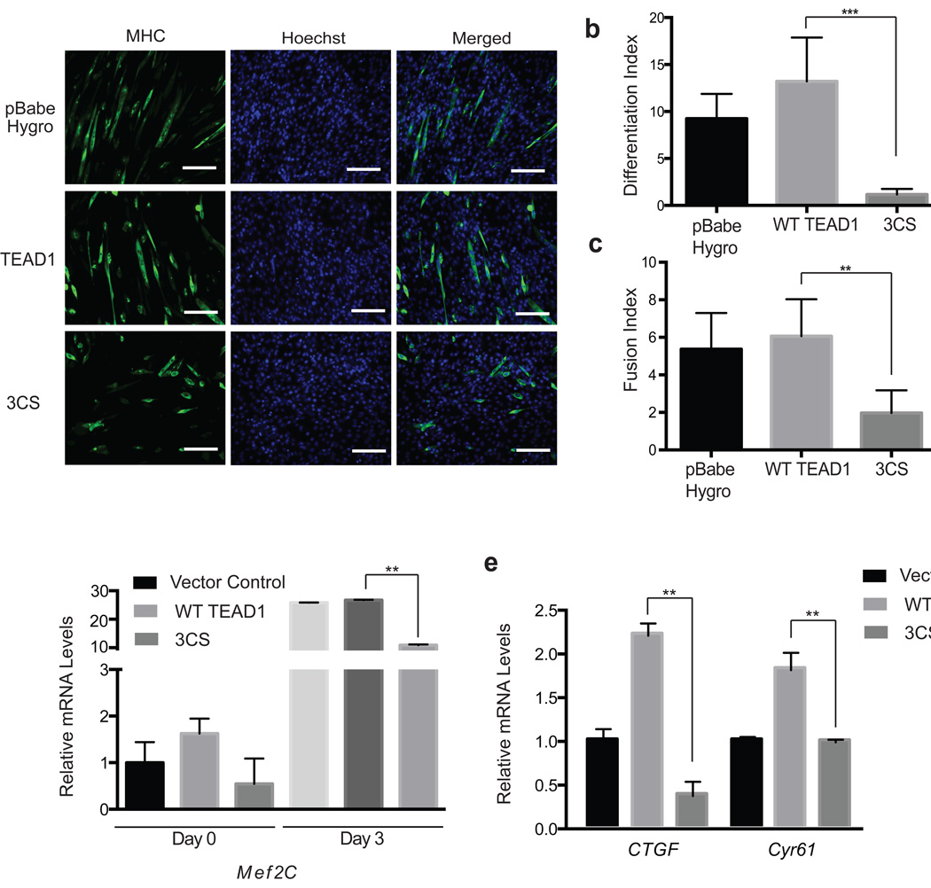
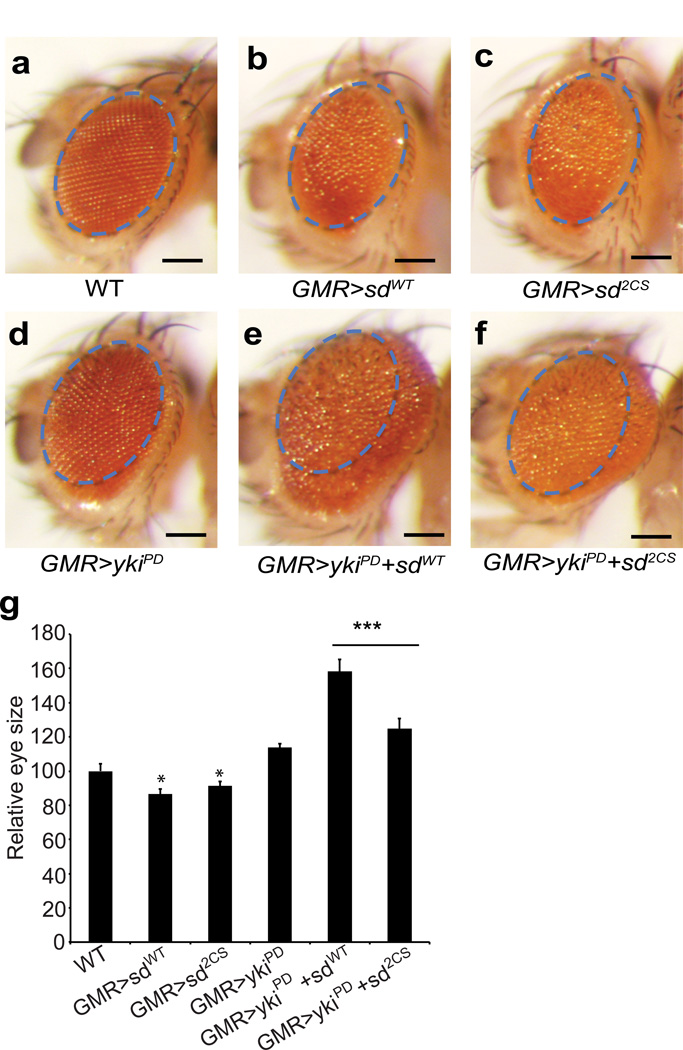
TEA domain (TEAD) transcription factors bind to the coactivators YAP and TAZ and regulate the transcriptional output of the Hippo pathway, playing critical roles in organ size control and tumorigenesis. Protein S-palmitoylation attaches a fatty acid, palmitate, to cysteine residues and regulates protein trafficking, membrane localization and signaling activities. Using activity-based chemical probes, we discovered that human TEADs possess intrinsic palmitoylating enzyme-like activities and undergo autopalmitoylation at evolutionarily conserved cysteine residues under physiological conditions. We determined the crystal structures of lipid-bound TEADs and found that the lipid chain of palmitate inserts into a conserved deep hydrophobic pocket. Strikingly, palmitoylation did not alter TEAD's localization, but it was required for TEAD's binding to YAP and TAZ and was dispensable for its binding to the Vgll4 tumor suppressor. Moreover, palmitoylation-deficient TEAD mutants impaired TAZ-mediated muscle differentiation in vitro and tissue overgrowth mediated by the Drosophila YAP homolog Yorkie in vivo. Our study directly links autopalmitoylation to the transcriptional regulation of the Hippo pathway.


Figures

Figure 1
Figure 1. Chemical approaches reveal that TEA domain (TEAD) transcription factors are palmitoylated
(a) Structures of the chemical reporter of palmitoylation (1), and the activity-based chemical probes for palmitoyl acyltransferases (PATs) and autopalmitoylated proteins (2 and 3). (b) 1 and 2 labeled myc-TEAD1 and myc-TEAD4 in HEK293A cells. The streptavidin blot showed the palmitoylation of TEADs. (c) Endogenous human TEAD1– 4 are all palmitoylated. The palmitoylated proteome of HEK293A and MCF10A cells was labeled by 1, and enriched by streptavidin beads. Western blots of TEAD1– 4 were carried out in the pull-down samples using anti-TEAD1, 2, 3, 4 antibodies. See Supplementary Fig. 10 for the full image of the blots. (d) TEAD1 is S-palmitoylated and hydroxylamine treatment dramatically decreased its palmitoylation levels. See Supplementary Fig. 10 for the full image of the blots.
Figure 2
Figure 2. TEAD is autopalmitoylated at evolutionarily conserved cysteine residues under physiological palmitoyl-CoA concentrations
(a) Mutation of the conserved cysteine residues (C53, C327 and C359) to serine residues individually or in combination (b) blocked palmitoylation of TEAD1. See Supplementary Figure 11 for the full image of the blots. (c) Recombinant TEAD2 protein (YAP binding domain, YBD) is autopalmitoylated in vitro in the presence of alkyne palmitoyl-CoA. See Supplementary Fig. 11 for the full image of the blots. (d) Mass spectrometry analysis of recombinant TEAD2 YBD reveals palmitoylation of TEAD2. (e) Acyl-biotin exchange (ABE) assay confirmed autopalmitoylation of recombinant TEAD2 YBD. See Supplementary Fig. 11 for the full image of the blots. (f) The Km value of palmitoyl-CoA in TEAD2 autopalmitoylation was estimated by plotting the reaction rate against the substrate concentration.
Figure 3
Figure 3. Structures of palmitate-bound human TEAD2 YBD and TEAD1–YAP complex
The Fo – Fc omit electron density map for TEAD2 (a) and TEAD1–YAP (c) at the contour level of 2.5σ. Palmitate (PLM) is shown as yellow sticks, and surrounding residues are shown as cyan sticks. Palmitate is covalently linked to C359 of TEAD1 (c). Ribbon diagram (left) and electrostatic surface (right) of PLM-bound TEAD2 YBD (PDB code: 5HGU) (b) and TEAD1–YAP complex (d) are shown. TEADs are colored in cyan and YAP is colored in pink. Two conserved cysteine residues are shown. The surface opening in free TEAD2 and the corresponding position in TEAD1–YAP are indicated by red arrow. All structural figures were generated with PyMOL (https://www.pymol.org).
Figure 4
Figure 4. Palmitoylation of TEAD is required for its association with YAP/TAZ
(a) Palmitoylation-deficient mutants of TEAD1 (C359S, C327/359S (2CS), and 3CS) have decreased association with YAP in co-immunoprecipitation (co-IP) experiments. See Supplementary Fig. 12 for the full image of the blots. (b) YAP binds to and significantly activates Gal4-TEAD1 wild type (WT) in Gal4-responsive luciferase assay. The palmitoylation-deficient Gal4-TEAD1 mutants (C359S, 2CS and 3CS) significantly inhibits Gal4-responsive luciferase reporter. (Data are represented as mean ± SEM, n=3. P values were determined using two-tailed t-tests. ****, p<0.0001, **, p<0.005). (c) FRET-based binding assay (Alpha Screen) showed that TEAD1 palmitoylation-deficient mutants (C359S, 2CS and 3CS) have decreased binding to YAP, comparing to TEAD1 WT. (Data are represented as mean ± SEM, n=3. P values were determined using two-tailed t-tests. ***, p<0.0005). (d) Palmitoylation-deficient mutants of TEAD1 (C359S, 2CS, and 3CS) significantly decreased TEAD transcription activity shown in a TEAD-binding element driven luciferase reporter assay (8XGTIIC-luciferase). (Data are represented as mean ± SEM, n=3. P values were determined using two-tailed t-tests. *, p<0.05; **, p<0.005). (e) Palmitoylation-deficient mutants of TEAD1 (C359S, 2CS, and 3CS) retain the binding to Vgll4 tumor suppressor in co-immunoprecipitation (co-IP) experiments. See Supplementary Fig. 12 for the full image of the blots. (f) FRET-based binding assay (Alpha Screen) showed that TEAD1 palmitoylation-deficient mutants (C359S, 2CS and 3CS) and TEAD1 WT bind to Vgll4 similarly. (Data are represented as mean ± SEM, n=3. P values were determined using two-tailed t-tests. N.S., not significant)
Figure 5
Figure 5. Palmitoylation regulates TEAD functions in muscle cell differentiation in vitro
(a) Representative images of myosin heavy chain (MHC, green) immunostaining of C2C12 cells. C2C12 cells stably expressing vector control (pBabe Hygro), TEAD1 WT or TEAD1 3CS mutant, were induced to differentiate for 3 days. Cell nuclei were stained with DAPI (blue). Scale bar: 100µm. (b–c) TEAD1 3CS mutant significantly inhibited myogenic differentiation and myotube fusion. Differentiation and fusion indices were calculated by averaging the data obtained from five different fields. (Data are represented as mean ± SEM, n=5. P values were determined using two-tailed t-tests. **, p<0.005). (d–f) TEAD 3CS mutant blocked the expression of myogenic markers Mef2C, and TEAD target genes (CTGF and Cyr61) in C2C12 cell. RNA samples of C2C12 stably expressed vector control, wild type and 3CS mutant of TEAD1 were collected and cDNA of each were synthesized. mRNA levels of each gene were determined by qRT-PCR using SYBR Green and normalized to GAPDH. (Data are represented as mean ± SEM, n=3. P values were determined using two-tailed t-tests. **, p<0.01)
Figure 6
Figure 6. Palmitoylation is required for the functions of Drosophila TEAD protein Scalloped (Sd) in vivo
Images of compound eyes from the following genotypes: (a) GMR-gal4/+, (b) GMR-gal4/UAS-sdWT, (c) GMR-gal4/UAS-sd2CS, (d) GMR-gal4, UAS-ykiPD, (e) GMR-gal4, UAS-ykiPD/UAS-sdWT, (f) GMR-gal4, UAS-ykiPD/UAS-sd2CS. Scale bar: 150µm. Note the overgrowth phenotype (enlarged eyes with rough surface) caused by co-expression of Yki-PD and Sd (WT) (e) is compromised when Yki-PD is co-expressed with the palmitoylation-deficient Sd (2CS) mutant (f). The images were taken with the same magnification. The size of the eye in wild type control flies is marked in blue dashed line, and the same area is shown in all images to facilitate comparison across all genotypes. (g) Relative sizes of the fly eyes are quantified in indicated genotypes. Sd WT and Sd 2CS flies are compared to the wild type, with statistically smaller eyes. (Data are represented as mean ± SEM, n=10 for each genotype. P values were determined using two-tailed t-tests. *, p<0.05; ***, p<0.001)

References

    1. Harvey KF, Zhang X, Thomas DM. The Hippo pathway and human cancer. Nature reviews. Cancer. 2013;13:246–257. - PubMed
    1. Pan D. Hippo signaling in organ size control. Genes Dev. 2007;21:886–897. - PubMed
    1. Pan D. The hippo signaling pathway in development and cancer. Dev Cell. 2010;19:491–505. - PMC - PubMed
    1. Ota M, Sasaki H. Mammalian Tead proteins regulate cell proliferation and contact inhibition as transcriptional mediators of Hippo signaling. Development. 2008;135:4059–4069. - PubMed
    1. Wu S, Liu Y, Zheng Y, Dong J, Pan D. The TEAD/TEF family protein Scalloped mediates transcriptional output of the Hippo growth-regulatory pathway. Dev Cell. 2008;14:388–398. - PubMed

Publication types

MeSH terms

LinkOut - more resources