. 2016 Apr;19(4):623-33.
doi: 10.1038/nn.4256. Epub 2016 Feb 22.

Integrated genomics and proteomics define huntingtin CAG length-dependent networks in mice

Affiliations

Integrated genomics and proteomics define huntingtin CAG length-dependent networks in mice

Peter Langfelder et al. Nat Neurosci. 2016 Apr.

Abstract

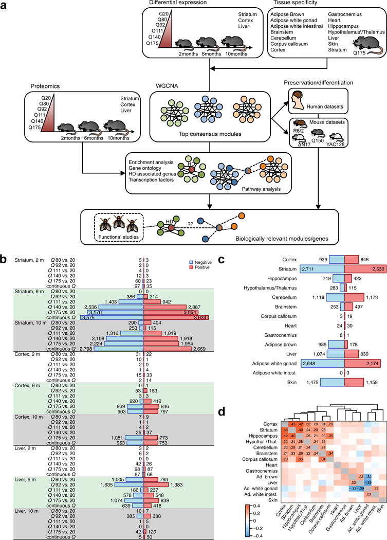
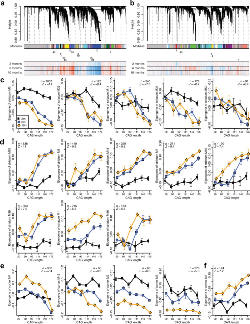
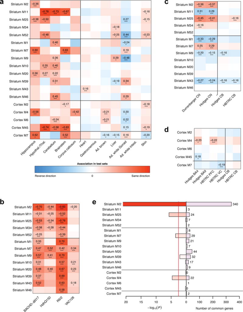
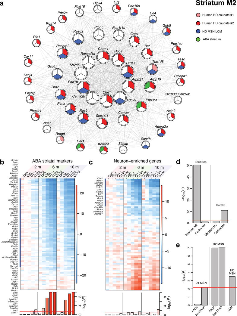
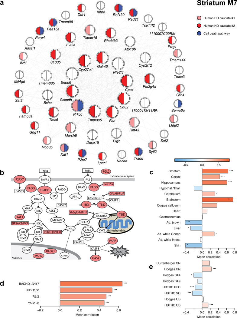
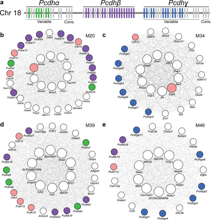
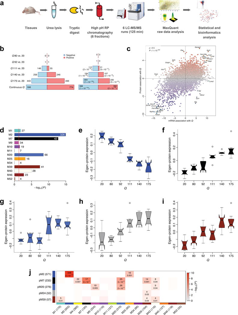
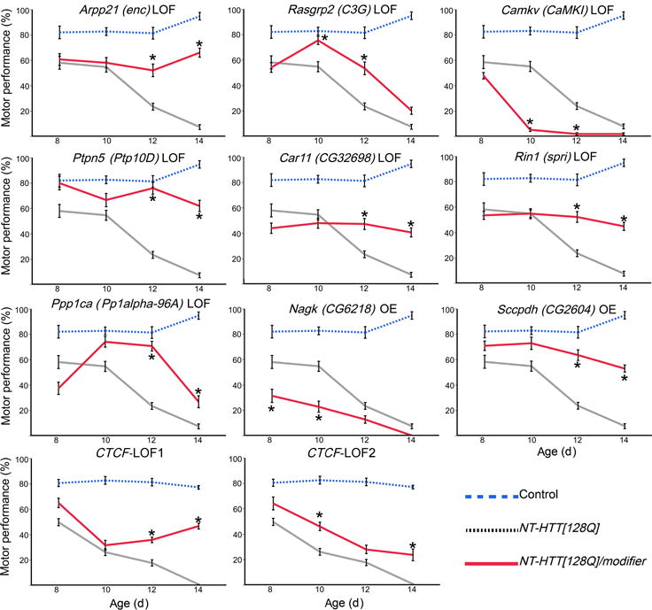
To gain insight into how mutant huntingtin (mHtt) CAG repeat length modifies Huntington's disease (HD) pathogenesis, we profiled mRNA in over 600 brain and peripheral tissue samples from HD knock-in mice with increasing CAG repeat lengths. We found repeat length-dependent transcriptional signatures to be prominent in the striatum, less so in cortex, and minimal in the liver. Coexpression network analyses revealed 13 striatal and 5 cortical modules that correlated highly with CAG length and age, and that were preserved in HD models and sometimes in patients. Top striatal modules implicated mHtt CAG length and age in graded impairment in the expression of identity genes for striatal medium spiny neurons and in dysregulation of cyclic AMP signaling, cell death and protocadherin genes. We used proteomics to confirm 790 genes and 5 striatal modules with CAG length-dependent dysregulation at the protein level, and validated 22 striatal module genes as modifiers of mHtt toxicities in vivo.


Conflict of interest statement

COMPETING FINANCIAL INTEREST

The authors declare that they have no competing interests that might be perceived to influence the results and/or discussion reported in this article.

Figures

Figure 1
Figure 1. Workflow and differential expression analysis with respect to Htt CAG length
(a) Overview of experiment design and analysis strategy. (b) Numbers of significantly (FDR<0.05) differentially expressed (DE) genes in striatum, cortex and liver. Blue (red) bars represent genes significantly down- (up-) regulated with increasing CAG length (Q). (c) Numbers of DE genes in the 14 tissues for which we profiled Q175 samples and controls including striatum, cortex, liver, and 11 additional tissues. The screening in the cortex and liver corresponds exactly to that presented in panel (b); the numbers for striatum are slightly different since the analysis in panel (b) used Q20 controls, while the one in panel (c) used WT controls. (d) Heatmap shows correlation of differential expression Z statistics of individual genes across the 14 data sets. Correlations whose absolute value is at least 0.2 are shown explicitly. Ad., adipose; intest., intestinal; Hypothal./Thal., hypothalamus/thalamus.
Figure 2
Figure 2. Consensus coexpression network analysis of striatum and cortex identifies multiple CAG length-dependent modules
(a and b) Striatum (a) and cortex (b) gene clustering trees. Each module (cluster) is labeled by a unique non-grey color, shown below the tree in the first color band, and a numeric label. For clarity, the numeric labels are shown only for modules that are strongly associated with CAG length Q (meta-analysis Z statistic |Z| > 5). The three-row heatmap below the tree indicates associations of individual genes with Q at each timepoint (2, 6, 10 months); blue and red indicate genes down- and up-regulated, respectively, with increasing Q. (c-f) Variation of module eigengenes with Q at each of the 3 ages (black: 2 months, blue: 6 months, orange: 10 months). Points represent means of eigengene “expression” across samples with a single CAG length. Error bars give SEM. Inset gives the number of genes in the module (n) and the meta-analysis association Z statistics for Q. Only modules with |Z| > 5 are shown. Eigengene expression values for the 6 month samples were shifted so that their mean equals the mean of the Q20 samples across 2 and 10 months. Panels (c) and (d) show eigengenes of striatum modules and (e) and (f) of cortex modules negatively and positively associated with Q, respectively.
Figure 3
Figure 3. Association of modules with genotype in mouse and disease status in human data and enrichment in human HD-dependent genes
(a-d) Weighted mean correlations (Methods) of module genes with genotype (mouse data) or HD status (human data). Statistical significance was determined from a permutation test and is indicated by the stars below each mean (*, P<0.05; **, P<0.01; ***, P<0.001; the p-values are listed explicitly in Supplementary Table S8). Red and blue color indicates the same and opposite direction, respectively, of differential expression in the discovery (striatum or cortex) and test set. (e) Enrichment of the CAG length-dependent striatal and cortical modules in genes that change consistently and significantly across human CN and cortex data (Methods). Bars show hypergeometric test p-values and numbers of common genes. Red line indicates Bonferroni-corrected threshold of 0.05. The enrichment p-value axis is truncated to 10−20 for clarity.
Figure 4
Figure 4. Striatal markers in module M2 undergo early and progressive CAG length-dependent changes
(a) Network of the top 50 hub genes in M2. Strong red, weak red or white color indicates significant change (FDR<0.1) in both, one of two or none of the human CN datasets; blue color indicates membership in top 100 ABA striatal markers; green indicates significant association with HD status in laser capture microdissection (LCM) data. (b, c) Heatmaps of striatum differential expression Z statistics of top ABA striatum neuronal marker genes (b) and ABA general neuronal marker genes (c). Genes that overlap with module M2 are marked with a blue bar and listed. Blue and red colors represent genes under- and over-expressed in higher CAG length compared to Q20, respectively. Bottom row shows the permutation test significance of the average of the Z statistics in each column. The y-axis range is restricted to 10 for clarity. Red line represents the significance threshold P=0.05. (d) Hypergeometric enrichment p-values of striatum module M2 and cortex module M4 in top ABA striatal and cortical markers. Red line indicates the Bonferroni-corrected threshold of 0.05. (e) Hypergeometric enrichment p-values of striatum module M2 in D1- and D2-MSN specific genes determined by FACS and bacTRAP, and in genes significantly associated with HD status in LCM data.
Figure 5
Figure 5. Cell death genes in striatum module M7
(a) Network plot of the top 50 hub genes in striatum module M7. Strong red, weak red and white color indicate significant (FDR<0.1) and consistent DE between HD samples and controls in both, one or none of the human CN datasets; blue color indicates genes implicated in cell death. (b) Graphical representation of the cell death pathway with dysregulated genes from striatum module M7 highlighted in red. (c-e) Association of cell death genes in striatum module M7 with genotype or HD status in other mouse striatum data (c), the tissue survey (d), and human post-mortem data (e). Bars show the weighted average correlation of the cell death M7 genes with the relevant genotype or HD status; stars show the corresponding permutation test significance (*, P<0.05; **, P<0.01; ***, P<0.001). The correlations and p-values are also listed in Supplementary Table S12.
Figure 6
Figure 6. Protocadherin dysregulation across multiple modules
(a) Schematic of the clustering of mouse protocadherins. Each vertical bar represents one of the protocadherin genes. Colored bars show protocadherins that are members of CAG length-dependent allelic series consensus modules. (b-e) Network plots of top 10 hub genes (center ring), and other hub genes and protocadherins (outer ring) in striatal modules M20 (b), M34 (c), M39 (d), and M46 (e). Colored circles indicate clustered (colors corresponding to schematic above: green, Pcdhα; purple Pcdhβ; blue, Pcdhγ) and unclustered protocadherins (pink).
Figure 7
Figure 7. High-throughput proteomic analysis confirms CAG length-dependent changes in 6-month striatum of HD mice
(a) Workflow of proteomic profiling. (b) Left barplot represents numbers of proteins significantly (FDR<0.1) associated with CAG length (Q). Blue (red) bars denote proteins down- (up-) regulated with increasing CAG length. Right barplot represents numbers of significant proteins whose mRNA also changes significantly (FDR<0.1) and in the same direction. Values in brackets are hypergeometric p-values of overlaps of significant protein and gene mRNA profiles. (c) Z statistics for protein association with Q vs. the corresponding mRNA Z statistics. Blue (red) dots represent proteins whose abundance decreases (increases) with increasing CAG length. Selected concordant and discordant genes are labeled. (d) Bars show hypergeometric enrichment p-values of mRNA module genes in proteins that are significantly differentially abundant in the same direction as the module. Numbers give the corresponding gene counts. For clarity, the p-value axis is truncated to 10−15. (e-i) Summary profiles of the 5 protein network modules with strongest association with Q, as a function of Q. Boxes indicate the median, interquartile range and confidence interval for the median. Whiskers indicate the range of data up to 1.5 of the inter-quartile range; points beyond the range of whiskers (if any) are shown individually. (j). Numbers of common genes and hypergeometric overlap p-values among selected protein (rows) and mRNA (columns) modules. Row and column labels indicate module sizes within the 7,039 genes common to both mRNA and protein network analyses.
Figure 8
Figure 8. Genetic perturbation studies in a fly model expressing mHTT fragment
Summary of motor performance tests for selected modifiers. Blue line represents controls, black line represents transgenic (NT-HTT[128Q]) flies, and red line represents transgenic flies with an active modifier. Each data point represents two replicates of 15 age-matched females per genotype and 10 trials (n=20). Error bars indicate SEM. Dunnett’s test following ANOVA was used to quantify statistical significance. Supplementary Figure 6 lists the relevant test statistics (ANOVA F values and Dunnett’s test p-values). Stars indicate pairs of means that are significantly different between NT-HTT[128Q] and NT-HTT[128Q]/modifier (p<0.05).

References

    1. Ross CA, et al. Huntington disease: natural history, biomarkers and prospects for therapeutics. Nature reviews. Neurology. 2014;10:204–216. - PubMed
    1. Vonsattel JP, DiFiglia M. Huntington disease. Journal of neuropathology and experimental neurology. 1998;57:369–384. - PubMed
    1. A novel gene containing a trinucleotide repeat that is expanded and unstable on Huntington’s disease chromosomes. The Huntington’s Disease Collaborative Research Group. Cell. 1993;72:971–983. - PubMed
    1. Orr HT, Zoghbi HY. Trinucleotide repeat disorders. Annual review of neuroscience. 2007;30:575–621. - PubMed
    1. Gusella JF, MacDonald ME. Molecular genetics: unmasking polyglutamine triggers in neurodegenerative disease. Nature reviews. Neuroscience. 2000;1:109–115. - PubMed

Publication types