Randomized Controlled Trial
. 2016 Feb 22;11(2):e0149219.
doi: 10.1371/journal.pone.0149219. eCollection 2016.

Dynamic Drusen Remodelling in Participants of the Nutritional AMD Treatment-2 (NAT-2) Randomized Trial

Affiliations
Randomized Controlled Trial

Dynamic Drusen Remodelling in Participants of the Nutritional AMD Treatment-2 (NAT-2) Randomized Trial

Giuseppe Querques et al. PLoS One. .

Abstract

Purpose: To evaluate the dynamic remodeling of drusen in subjects with unilateral neovascular age-related macular degeneration (AMD) receiving a three-year course of oral docosahexaenoic acid (DHA) or placebo.

Setting: Institutional setting.

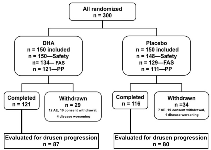
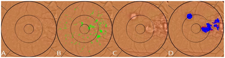
Methods: Three hundred subjects with age-related maculopathy and neovascular AMD in the fellow eye were randomly assigned to receive either 840 mg/day DHA or placebo for 3 years. Main outcome measures of this post-hoc sub-group analysis were progression of drusen number, total diameter, and total area on fundus photography, and their association with DHA supplementation, socio-demographic and genetic characteristics.

Results: Drusen progression was analyzed in 167 subjects that did not develop CNV (87 that received DHA and 80 that received placebo). None of the drusen remodeling outcomes were significantly associated with DHA supplementation. Total drusen diameter reduction in the inner subfield was significantly associated with age (older patients: r = -0.17; p = 0.003). Women showed a tendency to decreased total drusen diameter in the inner subfield with CFH polymorphism (p = 0.03), where women with TT genotype tended to have a greater reduction in drusen diameter than other genotypes (CC and CT). Drusen area in the inner subfield was more reduced in older patients (r = -0.17) and in women (p = 0.01). Drusen number showed no significant trends.

Conclusions: Dynamic drusen remodeling with net reduction in drusen load over three years was found in patients with exudative AMD in one eye and drusen in the other eye (study-eye). This reduction was correlated with increased age and female gender, and showed a tendency to be influenced by CFH genotype, but did not appear to be affected by DHA supplementation.

Trial registration: Controlled-Trials.com ISRCTN98246501.


Conflict of interest statement

Competing Interests: This study was supported by a grant from Laboratoire Bausch & Lomb - Chauvin, Clinical Research, 416, rue Samuel Morse, CS 99535, 34961 Montpellier, Cedex 2, France. Dr Querques is a consultant for Novartis, Bayer, Allergan, has received travel expenses from Novartis, Bayer, Allergan. Dr Merle has received consulting support from Bausch & Lomb, grants from Fédération des Aveugles et Handicapés Visuels de France and Fondation Nestlé France. Dr Benlian has received consulting and lecture support from Bausch & Lomb - Chauvin, Novartis. CD: Bausch & Lomb - Chauvin (consulting, conferences), Laboratoires Théa (Clermont-Ferrand, France) (research funding); Alcon (conferences). Dr Souied is a consultant for Novartis, Bayer, Allergan, has received travel expenses from Novartis, Bayer, Allergan and serves on the Speakers' Bureau of Novartis. There are no patents, products in development or marketed products to declare. This does not alter the authors’ adherence to all the PLOS ONE policies on sharing data and materials, as detailed online in the guide for authors.

Figures

Fig 1
Fig 1. Diagram showing the study population.
The safety population included all subjects who have received the study treatment. Full analysis set (FAS) included all subjects in the safety set who have had at least 1 post-baseline evaluation regarding occurrence of choroidal neovascularization. Per protocol (PP) population included all FAS subjects without major protocol deviation. Among those withdrawn there were 3 deaths in the docosahexaenoic acid (DHA) group and 6 deaths in the placebo group.
Fig 2
Fig 2. Illustration of drusen segmentation methods.
A) Red-free fundus photo. Areas with drusen are circled in black (human supervision) to initialize the segmentation program. This allows the program to exclude from consideration peripapillary changes and other hypo-pigmented or hyper-reflective non-drusen features, resulting in a more rapid and accurate drusen identification. B) original color fundus photograph, cropped to 6 mm field C) contrast-enhanced color photograph D) contrast-enhanced color photograph with superimposed Wisconsin grading template (6 mm diameter circle with central, middle and outer subfields of diameters of 1, 3 and 6 mm). Drusen have been automatically segmented in detail (green) by the custom software within the areas containing drusen identified in A.
Fig 3
Fig 3. Segmentation of reticular pseudodrusen.
A) original fundus photograph with large area containing both reticular macular disease (reticular pseudodrusen) and ordinary soft drusen outlined in black. The soft drusen are confined to the central macula. The reticular pseudodrusen extend to the arcades. B) original color fundus photo C) both reticular pseudodrusen and soft drusen are segmented (green) on the color photo within the area identified in A and within the superimposed Wisconsin grading template. D) The area of reticular macular disease is separately enclosed in red, and was excluded from drusen measurements.
Fig 4
Fig 4. Segmentation of drusen and geographic atrophy (GA).
A) Original red-free fundus photograph B) Original color fundus photograph with areas of drusen and GA outlined (black) C) Color fundus photograph cropped to 6 mm field. D) contrast enhanced color photo. E) segmentation of drusen (green) within the 6 mm field F) areas of GA masked (black).
Fig 5
Fig 5. Drusen regression and its correlation with new geographic atrophy (GA).
A: Color photo, right eye, Visit 1 (V1). B: V1 photo with soft drusen segmented in green, total pixel area 2,452. GA is not present. C: Color photo, right eye, Visit 5 (V5), showing new GA, D: V5 photo with GAs segmented in blue, total pixel area 4,161. Drusen from V1 that were absorbed in the GA are overlaid and segmented in red, total pixel area 381, or 9% of geographic atrophy total area. Compared to the original drusen area present in V1 of 2,452 pixels, the 381 drusen pixels converting to GA in V5 represent 16% of the original drusen area.

References

    1. Resnikoff S, Pascolini D, Etya'ale D, Kocur I, Pararajasegaram R, Pokharel GP, et al. Global data on visual impairment in the year 2002. Bull World Health Organ. 2004; 82(11):844–851. - PMC - PubMed
    1. Ferris FL, Davis MD, Clemons TE, Lee LY, Chew EY, Lindblad AS, et al. A simplified severity scale for age-related macular degeneration: AREDS Report No. 18. Arch Ophthalmol. 2005;123(11):1570–1574. - PMC - PubMed
    1. Seddon JM, Sharma S, Adelman RA. Evaluation of the clinical age-related maculopathy staging system. Ophthalmology. 2006;113(2):260–266. - PubMed
    1. Pauleikhoff D, Barondes MJ, Minassian D, Chisholm IH, Bird AC. Drusen as risk factors in age-related macular disease. Am J Ophthalmol.1990;109(1): 38–43. - PubMed
    1. Klein R, Klein BE, Knudtson MD, Meuer SM, Swift M, Gangnon RE. Fifteen-year cumulative incidence of age-related macular degeneration: the Beaver Dam Eye Study. Ophthalmology. 2007;114(2): 253–262. - PubMed

Publication types

Substances

Associated data