Clinical Trial
. 2016 Feb 22;11(2):e0149770.
doi: 10.1371/journal.pone.0149770. eCollection 2016.

High Resolution Topography of Age-Related Changes in Non-Rapid Eye Movement Sleep Electroencephalography

Affiliations
Clinical Trial

High Resolution Topography of Age-Related Changes in Non-Rapid Eye Movement Sleep Electroencephalography

Kate E Sprecher et al. PLoS One. .

Abstract

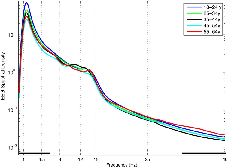
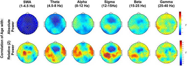
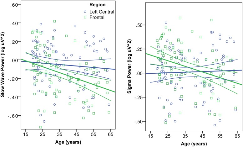
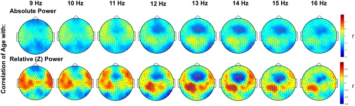
Sleeping brain activity reflects brain anatomy and physiology. The aim of this study was to use high density (256 channel) electroencephalography (EEG) during sleep to characterize topographic changes in sleep EEG power across normal aging, with high spatial resolution. Sleep was evaluated in 92 healthy adults aged 18-65 years old using full polysomnography and high density EEG. After artifact removal, spectral power density was calculated for standard frequency bands for all channels, averaged across the NREM periods of the first 3 sleep cycles. To quantify topographic changes with age, maps were generated of the Pearson's coefficient of the correlation between power and age at each electrode. Significant correlations were determined by statistical non-parametric mapping. Absolute slow wave power declined significantly with increasing age across the entire scalp, whereas declines in theta and sigma power were significant only in frontal regions. Power in fast spindle frequencies declined significantly with increasing age frontally, whereas absolute power of slow spindle frequencies showed no significant change with age. When EEG power was normalized across the scalp, a left centro-parietal region showed significantly less age-related decline in power than the rest of the scalp. This partial preservation was particularly significant in the slow wave and sigma bands. The effect of age on sleep EEG varies substantially by region and frequency band. This non-uniformity should inform the design of future investigations of aging and sleep. This study provides normative data on the effect of age on sleep EEG topography, and provides a basis from which to explore the mechanisms of normal aging as well as neurodegenerative disorders for which age is a risk factor.


Conflict of interest statement

Competing Interests: Giulio Tononi has consulted for Philips Respironics and has been involved in a research study in humans supported by Philips Respironics. Giulio Tononi is also a consultant for the Allen Institute for Brain Research. Ruth M. Benca has served as a consultant to Merck and Jazz and received grant support from Merck. The article submitted is not related to any of these relationships. The remaining authors have reported that no competing interests exist. This did not alter the authors' adherence to PLOS ONE policies on sharing data and materials.

Figures

Fig 1
Fig 1. NREM EEG Power Spectra.
EEG power spectra (log μV2/Hz) for NREM sleep during the first 3 sleep cycles, averaged across 173 scalp electrodes in 1/6 Hz frequency bins. Age groups plotted by decade (aged 18–25 years, dark blue; 25–35 years, green; 35–45 years, black; 45–55 years, light blue; 55–65 years, red). Classically defined frequency bands indicated by vertical dotted lines. Black squares along the x-axis indicate frequency bins in which ANOVA showed a significant effect of age group.
Fig 2
Fig 2. Topography of the correlation of age with NREM EEG power.
Topography of the correlation between age and NREM spectral density, averaged across the first three sleep cycles in standard frequency bands. Color represents the coefficient of the correlation between age and power at each electrode. In the upper panel, blue indicates a negative correlation, i.e. a decline in absolute NREM EEG power (log μV2) with increasing age. The lower panel plots the coefficient of correlation (r) between age and power (μV2) normalized to the scalp mean within an individual. Colors indicate that as age increases, EEG power is increasingly higher (red) or lower (blue) than the scalp mean. Large black dots indicate channels at which the correlation of age and EEG power was significant, accounting for multiple comparisons with statistical nonparametric mapping.
Fig 3
Fig 3. Interaction of age and region on EEG Power.
Correlation of age and absolute NREM EEG power (log μV2) in the Slow Wave (left) and Sigma (right) frequency bands. In each band, power was averaged in a frontal (blue circles) and left central (green squares) region, defined by clusters of electrodes showing significant correlation of age and normalized power. Mixed ANOVA revealed a significant region x age interaction, such that the age-related decline of EEG power was greater in the frontal than left central region for slow wave and sigma bands.
Fig 4
Fig 4. Topography of the correlation of age with NREM EEG spectral density in spindle frequencies.
Topography of the correlation between age and NREM EEG spectral density (log μV2), in 1 Hz bins of spindle frequencies, averaged across the first three sleep cycles. Color represents the coefficient of the correlation between age and power at each electrode. Small black dots indicate electrode locations. Large black dots indicate electrodes at which the correlation of age and EEG power was significant, accounting for multiple comparisons with statistical nonparametric mapping.

References

    1. Buchmann A, Ringli M, Kurth S, Schaerer M, Geiger A, Jenni OG, et al. EEG Sleep Slow-Wave Activity as a Mirror of Cortical Maturation. Cereb Cortex. 2011;21: 607–615. 10.1093/cercor/bhq129 - DOI - PubMed
    1. Mander BA, Rao V, Lu B, Saletin JM, Lindquist JR, Ancoli-Israel S, et al. Prefrontal atrophy, disrupted NREM slow waves and impaired hippocampal-dependent memory in aging. Nat Neurosci. 2013;16: 357–364. 10.1038/nn.3324 - DOI - PMC - PubMed
    1. Dubé J, Lafortune M, Bedetti C, Bouchard M, Gagnon JF, Doyon J, et al. Cortical Thinning Explains Changes in Sleep Slow Waves during Adulthood. J Neurosci Off J Soc Neurosci. 2015;35: 7795–7807. 10.1523/JNEUROSCI.3956-14.2015 - DOI - PMC - PubMed
    1. Esser SK, Hill SL, Tononi G. Sleep Homeostasis and Cortical Synchronization: I. Modeling the Effects of Synaptic Strength on Sleep Slow Waves. Sleep. 2007;30: 1617–1630. - PMC - PubMed
    1. Vyazovskiy VV, Olcese U, Lazimy YM, Faraguna U, Esser SK, Williams JC, et al. Cortical firing and sleep homeostasis. Neuron. 2009;63: 865–878. 10.1016/j.neuron.2009.08.024 - DOI - PMC - PubMed

Publication types