. 2016 Jun;15(6):654-61.
doi: 10.1038/nmat4571. Epub 2016 Feb 22.

Lattice engineering through nanoparticle-DNA frameworks

Affiliations

Lattice engineering through nanoparticle-DNA frameworks

Ye Tian et al. Nat Mater. 2016 Jun.

Abstract

Advances in self-assembly over the past decade have demonstrated that nano- and microscale particles can be organized into a large diversity of ordered three-dimensional (3D) lattices. However, the ability to generate different desired lattice types from the same set of particles remains challenging. Here, we show that nanoparticles can be assembled into crystalline and open 3D frameworks by connecting them through designed DNA-based polyhedral frames. The geometrical shapes of the frames, combined with the DNA-assisted binding properties of their vertices, facilitate the well-defined topological connections between particles in accordance with frame geometry. With this strategy, different crystallographic lattices using the same particles can be assembled by introduction of the corresponding DNA polyhedral frames. This approach should facilitate the rational assembly of nanoscale lattices through the design of the unit cell.


Figures

Figure 1
Figure 1. Lattice assembly via Nanoparticle-DNA Framework
Nanoparticles (NPs) (yellow ball) capped with oligonucleotides (blue curves) are mixed with polyhedral DNA frames (from top to bottom): cube, octahedron, elongated square bipyramid (ESB), prism, triangular bipyramid (TBP). The frames are designed to have complementary strands at the vertices for NP binding. An aggregation (framework formation) is expected when NPs and the correspondingly encoded polyhedral frames are allowed to mix and hybridize. NP-DNA Frameworks might exhibit ordered lattice for optimized NP sizes, linking motif, and annealing process. The crystallographic symmetry of the assembled lattice is controlled by the shape of interparticle linking frames, but not by the NPs themselves. This strategy thus enables “engineering” the lattice and its unit cell.
Figure 2
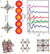
Figure 2. The formation of NP-DNA 3D lattice with octahedra frames and the effect of NP-vertex linker length
(a). The length of the linkers is increased (systems I to V, from bottom to the top) by changing the DNA linking motif. For respective systems (in parentheses) motifs are 0-6-10 (I), 2-6-10 (II), 9-6-10 (III), 6-9-21 (IV), 0-15-15 (V), see also Table 1 and Supplementary Information. The central panel shows 2D SAXS data for each system. The corresponding structure factors S(q/q0) are shown (coloured lines/circles) on the right. The indexing of scattering peaks positions and intensities for FCC lattice is shown (black lines). (b). Proposed assembled superlattice of NPs and octahedra. The particles within a single unit cell (marked by red lines) of FCC structure are shown in red on the left panel. The corresponding side and top views of the FCC structured NP-DNA framework are shown on the centre and right panels respectively.
Figure 3
Figure 3. Nanoparticle superlattices assembled via DNA frames
(a – d) DNA-coated NPs (10 nm gold core) bound to four different types of frames via complementary DNA strands at their respective vertices. The four polyhedral frames are: octahedron (a1), ESB (b1), cube (c1) and prism (d1). For each, we show EM image (2) (see the Supplementary Information for the details of 3D reconstruction from cryo-TEM measurements), x-ray scattering structure factor, S(q) with the 2D SAXS pattern as inset (3) were obtained in-situ after the annealing of the formed NP-DNA frameworks, and the proposed superlattice structures, as discussed in the text. See Supplementary Fig. 14–17 for the enlarged views of NP lattice models and the idealized arrangements of DNA frames for the obtained frames to NPs ratios, see the text (4). For each lattice structure, the experimental scattering profile is in blue and the model fitting in red. The black peaks mark the standard peak positions of the proposed lattice: FCC by the octahedra (a3), BCT by ESB (b3), simple cubic by the cubes (c3), and hexagonal by the prisms (d3).
Figure 4
Figure 4. Cryo-STEM images for simple cubic (SC) (a1, a2) and body centred tetragonal (BCT) (b1, b2) lattices of NPs assembled with cubic and ESB frames, respectively
(a1) STEM image of the (111) plane projection for the SC lattice; the nearest interparticle distance is 40.2nm. (a2) STEM image of the (100) face projection for the SC lattice which is tilted in one direction; the nearest interparticle distance is 50.7nm. (b1) STEM image of the (111) face projection of the BCT lattice, the nearest interparticle distance is 46.5nm. (b2) STEM image of the (100) face projection of the BCT lattice, the nearest interparticle distance is 53.2nm. The top left and right parts of each panel display the lattice model with the corresponding projection and the particle arrangements, respectively. The corresponding particles from the models and images are highlighted by red circles.

References

    1. Talapin DV, Lee J-S, Kovalenko MV, Shevchenko EV. Prospects of Colloidal Nanocrystals for Electronic and Optoelectronic Applications. Chem. Rev. 2010;110:389–458. - PubMed
    1. Hu T, Isaacoff BP, Bahng JH, Hao C, Zhou Y, Zhu J, Li X, Wang Z, Liu S, Xu C, Biteen JS, Kotov NA. Self-Organization of Plasmonic and Excitonic Nanoparticles into Resonant Chiral Supraparticle Assemblies. Nano Lett. 2014;14:6799–6810. - PubMed
    1. Cargnello M, Johnston-Peck AC, Diroll BT, Wong E, Datta B, Damodhar D, Doan-Nguyen VVT, Herzing AA, Kagan CR, Murray CB. Substitutional doping in nanocrystal superlattices. Nature. 2015;524:450–453. - PubMed
    1. Kuzyk A, Schreiber R, Fan Z, Pardatscher G, Roller E-M, Högele A, Simmel FC, Govorov AO, Liedl T. DNA-based self-assembly of chiral plasmonic nanostructures with tailored optical response. Nature. 2012;483:311–314. - PubMed
    1. Xiong H, Sfeir MY, Gang O. Assembly, structure and optical response of three-dimensional dynamically tunable multicomponent superlattices. Nano Lett. 2010;10:4456–4462. - PubMed

Publication types

MeSH terms