Rho Kinase ROCK2 Mediates Acid-Induced NADPH Oxidase NOX5-S Expression in Human Esophageal Adenocarcinoma Cells
- PMID: 26901778
- PMCID: PMC4764682
- DOI: 10.1371/journal.pone.0149735
Rho Kinase ROCK2 Mediates Acid-Induced NADPH Oxidase NOX5-S Expression in Human Esophageal Adenocarcinoma Cells
Abstract
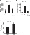
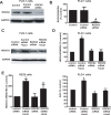
Mechanisms of the progression from Barrett's esophagus (BE) to esophageal adenocarcinoma (EA) are not fully understood. We have shown that NOX5-S may be involved in this progression. However, how acid upregulates NOX5-S is not well known. We found that acid-induced increase in NOX5-S expression was significantly decreased by the Rho kinase (ROCK) inhibitor Y27632 in BE mucosal biopsies and FLO-1 EA cells. In addition, acid treatment significantly increased the Rho kinase activity in FLO-1 cells. The acid-induced increase in NOX5-S expression and H2O2 production was significantly decreased by knockdown of Rho kinase ROCK2, but not by knockdown of ROCK1. Conversely, the overexpression of the constitutively active ROCK2, but not the constitutively active ROCK1, significantly enhanced the NOX5-S expression and H2O2 production. Moreover, the acid-induced increase in Rho kinase activity and in NOX5-S mRNA expression was blocked by the removal of calcium in both FLO-1 and OE33 cells. The calcium ionophore A23187 significantly increased the Rho kinase activity and NOX5-S mRNA expression. We conclude that acid-induced increase in NOX5-S expression and H2O2 production may depend on the activation of ROCK2, but not ROCK1, in EA cells. The acid-induced activation of Rho kinase may be mediated by the intracellular calcium increase. It is possible that persistent acid reflux present in BE patients may increase the intracellular calcium, activate ROCK2 and thereby upregulate NOX5-S. High levels of reactive oxygen species derived from NOX5-S may cause DNA damage and thereby contribute to the progression from BE to EA.
Conflict of interest statement
Figures


Similar articles
-
Role of intracellular calcium and NADPH oxidase NOX5-S in acid-induced DNA damage in Barrett's cells and Barrett's esophageal adenocarcinoma cells.Am J Physiol Gastrointest Liver Physiol. 2014 May 15;306(10):G863-72. doi: 10.1152/ajpgi.00321.2013. Epub 2014 Apr 3. Am J Physiol Gastrointest Liver Physiol. 2014. PMID: 24699332 Free PMC article.
-
Role of Rac1 in regulation of NOX5-S function in Barrett's esophageal adenocarcinoma cells.Am J Physiol Cell Physiol. 2011 Aug;301(2):C413-20. doi: 10.1152/ajpcell.00027.2011. Epub 2011 Apr 27. Am J Physiol Cell Physiol. 2011. PMID: 21525435 Free PMC article.
-
NADPH oxidase NOX5-S mediates acid-induced cyclooxygenase-2 expression via activation of NF-kappaB in Barrett's esophageal adenocarcinoma cells.J Biol Chem. 2007 Jun 1;282(22):16244-55. doi: 10.1074/jbc.M700297200. Epub 2007 Apr 2. J Biol Chem. 2007. PMID: 17403674
-
Isoform-specific targeting of ROCK proteins in immune cells.Small GTPases. 2016 Jul 2;7(3):173-7. doi: 10.1080/21541248.2016.1181698. Epub 2016 Jun 2. Small GTPases. 2016. PMID: 27254302 Free PMC article. Review.
-
Distinct Roles of ROCK1 and ROCK2 on the Cerebral Ischemia Injury and Subsequently Neurodegenerative Changes.Pharmacology. 2020;105(1-2):3-8. doi: 10.1159/000502914. Epub 2019 Sep 19. Pharmacology. 2020. PMID: 31537002 Review.
Cited by
-
Effect of Proton Pump Inhibitor Therapy on NOX5, mPGES1 and iNOS expression in Barrett's Esophagus.Sci Rep. 2019 Nov 7;9(1):16242. doi: 10.1038/s41598-019-52800-7. Sci Rep. 2019. PMID: 31700071 Free PMC article.
-
Vascular Biology of Superoxide-Generating NADPH Oxidase 5-Implications in Hypertension and Cardiovascular Disease.Antioxid Redox Signal. 2019 Mar 1;30(7):1027-1040. doi: 10.1089/ars.2018.7583. Epub 2018 Nov 15. Antioxid Redox Signal. 2019. PMID: 30334629 Free PMC article. Review.
-
The Impact of Oxidative Stress in Human Pathology: Focus on Gastrointestinal Disorders.Antioxidants (Basel). 2021 Jan 30;10(2):201. doi: 10.3390/antiox10020201. Antioxidants (Basel). 2021. PMID: 33573222 Free PMC article. Review.
-
TGR5 expression in benign, preneoplastic and neoplastic lesions of Barrett's esophagus: Case series and findings.World J Gastroenterol. 2017 Feb 28;23(8):1338-1344. doi: 10.3748/wjg.v23.i8.1338. World J Gastroenterol. 2017. PMID: 28293080 Free PMC article.
-
Carcinogenesis and Reactive Oxygen Species Signaling: Interaction of the NADPH Oxidase NOX1-5 and Superoxide Dismutase 1-3 Signal Transduction Pathways.Antioxid Redox Signal. 2019 Jan 20;30(3):443-486. doi: 10.1089/ars.2017.7268. Epub 2018 Nov 22. Antioxid Redox Signal. 2019. PMID: 29478325 Free PMC article. Review.
References
-
- Lagergren J, Bergstrom R, Lindgren A, Nyren O. Symptomatic gastroesophageal reflux as a risk factor for esophageal adenocarcinoma. N Engl J Med. 1999;340(11):825–31. . - PubMed
-
- Falk GW. Barrett's esophagus. Gastroenterology. 2002;122(6):1569–91. Epub 2002/05/23. S0016508502061620 [pii]. . - PubMed
-
- Ronkainen J, Aro P, Storskrubb T, Johansson SE, Lind T, Bolling-Sternevald E, et al. Prevalence of Barrett's esophagus in the general population: an endoscopic study. Gastroenterology. 2005;129(6):1825–31. . - PubMed
Publication types
MeSH terms
Substances
Supplementary concepts
Grants and funding
LinkOut - more resources
Full Text Sources
Other Literature Sources
Medical

