. 2016 Mar 1;126(3):1126-36.
doi: 10.1172/JCI84997. Epub 2016 Feb 22.

Neonatal thymectomy reveals differentiation and plasticity within human naive T cells

Neonatal thymectomy reveals differentiation and plasticity within human naive T cells

Theo van den Broek et al. J Clin Invest. .

Abstract

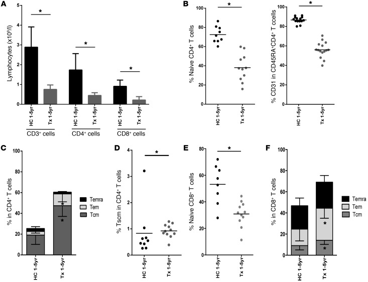
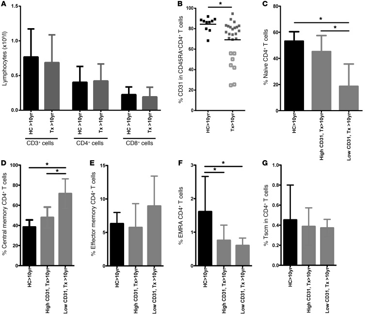
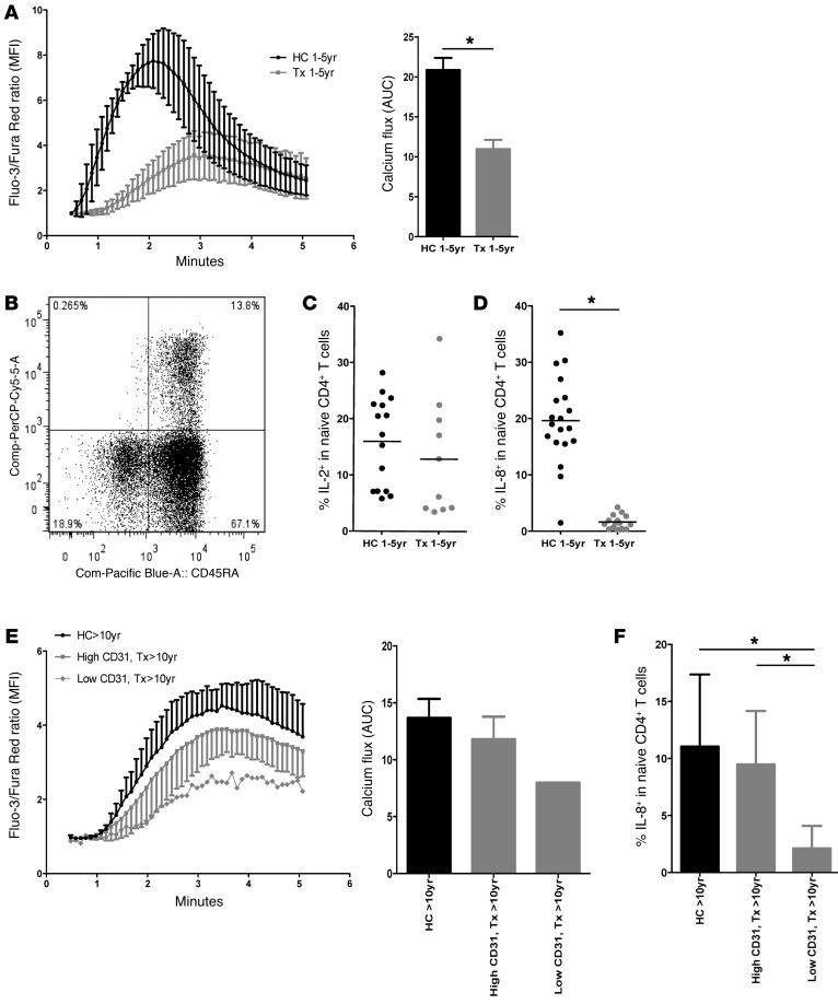
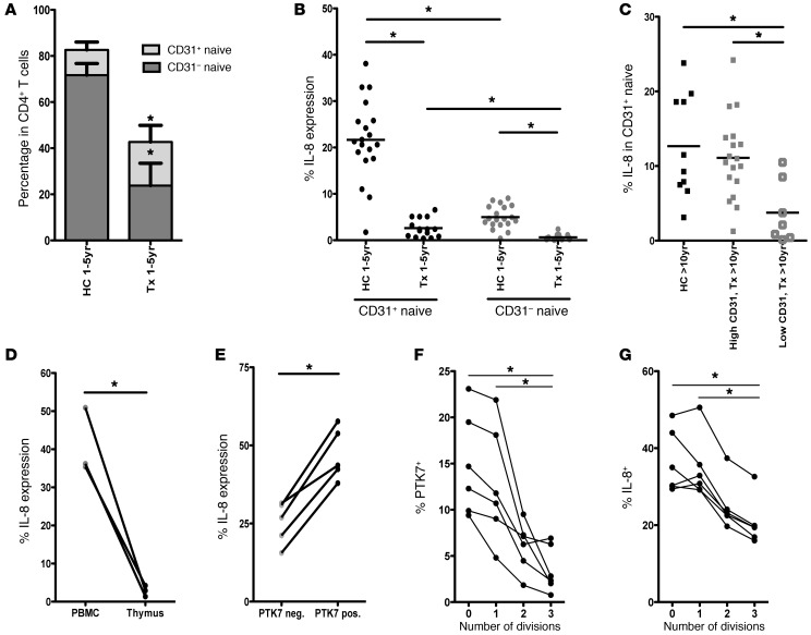
The generation of naive T cells is dependent on thymic output, but in adults, the naive T cell pool is primarily maintained by peripheral proliferation. Naive T cells have long been regarded as relatively quiescent cells; however, it was recently shown that IL-8 production is a signatory effector function of naive T cells, at least in newborns. How this functional signature relates to naive T cell dynamics and aging is unknown. Using a cohort of children and adolescents who underwent neonatal thymectomy, we demonstrate that the naive CD4+ T cell compartment in healthy humans is functionally heterogeneous and that this functional diversity is lost after neonatal thymectomy. Thymic tissue regeneration later in life resulted in functional restoration of the naive T cell compartment, implicating the thymus as having functional regenerative capacity. Together, these data shed further light on functional differentiation within the naive T cell compartment and the importance of the thymus in human naive T cell homeostasis and premature aging. In addition, these results affect and alter our current understanding on the identification of truly naive T cells and recent thymic emigrants.
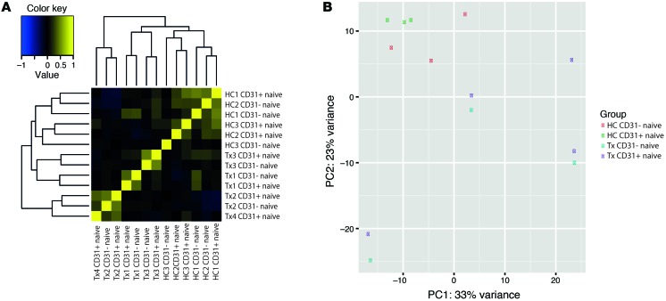

Figures

Figure 1
Figure 1. Neonatal thymectomy results in lower naive CD4+ T cell percentages and skewing toward a memory phenotype in the first years (1–5 years) of life.
(A) Blood lymphocyte count of CD3+, CD4+, and CD8+ T cells in HCs and neonatally Tx children for the age groups of 1 to 5 years. (B) Left panel, percentages of naive T cells (CD45RA+CCR7+) within the CD4+ T cell population. Right panel, percentages of CD31-expressing cells among CD45RA+CD4+ T cells. (C) Percentages of Tcm (CD45RACCR7+), Tem (CD45RACCR7), and Temra (CD45RA+CCR7) in the CD4+ T cell pool. (D) Percentages of Tscm (CD45RA+CCR7+CD28+CD27+FAS+) in the CD4+ T cell pool. (E) Percentages of naive T cells (CD45RA+CCR7+) within the CD8+ T cell population. (F) Percentages of Tcm (CD45RACCR7+), Tem (CD45RACCR7), and Temra (CD45RA+CCR7) in the CD8+ T cell pool. Black circles (or black bar, A), young HCs (n = 8–14); gray circles (gray bar, A), young Tx (n = 10–15). Data are shown as mean ± SD. See also Supplemental Figure 1. *P < 0.05, Mann-Whitney U test.
Figure 2
Figure 2. Thymic tissue regeneration results in restoration of the naive and effector T cell compartment later in life.
(A) Lymphocyte count of CD3+, CD4+, and CD8+ T cells in HCs and neonatally Tx children older than 10 years of age. (B) Percentages of CD31 expression on CD45RA+CD4+ T cells. (C) Percentages of naive T cells (CD45RA+CCR7+) within the CD4+ T cell population. (D) Percentages of Tcm (CD45RACCR7+). (E) Percentages of Tem (CD45RACCR7). (F) Percentages of Temra (CD45RA+CCR7). (G) Percentages of Tscm (CD45RA+CCR7+CD28+CD27+FAS+) in CD4+ T cells. Black squares (or black bars), older HCs (n = 10); gray squares (or gray bars), older Tx patients (n = 24–26). Older Tx patients are further divided in high percentages of CD31 (n = 17–19; closed gray square in B) and older TX with low CD31 percentages (n = 7; open gray squares in B), as described in the x axis. Data are shown as mean ± SD. See also Supplemental Figure 2. *P < 0.05, Mann-Whitney U test used.
Figure 3
Figure 3. Naive CD4+ T cell functionality is impaired after neonatal thymectomy in early life, but restores after thymic regeneration in later life.
(A) Left panel, calcium flux of CD4+ naive T cells in young children (young HCs, n = 4 black, young Tx, n = 6 gray, mean ± SD). Right panel, AUC of the calcium flux. (B) Representative dot plot of IL-8 production in CD45RA+CD4+ T cell population of young HCs. (C) IL-2 production in naive CD4+ T cells of young HCs (n = 15) and young Tx patients (n = 10) after PMA/ion stimulation. (D) IL-8 production by naive CD4+ T cells of young HC (n = 19) and young Tx patient (n = 15) PMA/ion stimulation. (E) Left panel, calcium flux of CD4 naive T cells of older HCs and Tx children. Right panel, AUC of the calcium flux (HC 1–5 yr, n = 5; high CD31, Tx > 10 yr, n = 6; low CD31, Tx > 10 yr, n = 1). (F) IL-8 production in naive CD4+ T cells of older HC (n = 10) and Tx patient (high CD31, n = 17; low CD31, n = 7) PMA/ion stimulation. Data are shown as mean ± SD. See also Supplemental Figure 3. *P < 0.05, Mann-Whitney U test.
Figure 4
Figure 4. IL-8 production is enriched in the PTK7+ fraction of CD31+ naive CD4+ T cells and lost after cell division.
(A) Proportion of CD31+ and CD31 naive CD4+ T cells in the CD4+ T cell compartment of HCs, 1–5 yr (n = 19) and Tx patients, 1–5 yr (n = 15). (B) Expression of IL-8 by CD31+ and CD31 naive CD4+ T cells of young HCs (n = 19) and Tx patients (n = 15). (C) Expression of IL-8 by CD31+ and CD31 naive CD4+ T cells of older HCs (n = 10) and older Tx patients separated on the basis of low (n = 7) or high percentage of CD31+ (n = 19). (D) Paired IL-8 expression measurements by SP CD3hiCD4+CD8 thymocytes and blood CD31+ naive CD4+ T cells (PBMCs) from the Tx neonates (n = 3). (E) IL-8 expression by PTK7+ (black dots) and PTK7 (gray dots) CD31+ naive CD4+ T cells from young HCs (n = 5). (F) PTK7 expression after each cell division following cytokine stimulation of FACS-sorted CD31+ naive CD4+ T cells from older HCs (n = 6). (G) IL-8 expression after each cell division following cytokine stimulation of FACS-sorted CD31+ naive CD4+ T cells from older HCs (n = 6). Data are shown as mean ± SD. See also Supplemental Figures 4 and 5. *P < 0.05, Mann-Whitney U test for unpaired data and Wilcoxon’s signed rank test for paired data.
Figure 5
Figure 5. Functional differentiation within CD31+ naive CD4+ T cells is lost after neonatal thymectomy.
(A) Correlation clustering of CD31+ and CD31 naive CD4+ T cells of young HCs (n = 3) and young Tx patients (n = 4). Yellow indicates high positive correlation, while blue indicates high negative correlation. (B) Principal component analysis of CD31+ and CD31 naive T cells of young HCs (n = 3) and young Tx patients (n = 4). See also Supplemental Figures 6 and 7 and Supplemental Tables 1–3.

Similar articles

Cited by

References

    1. Zhu J, Yamane H, Paul WE. Differentiation of effector CD4+ T cell populations. Annu Rev Immunol. 2010;28:445–489. doi: 10.1146/annurev-immunol-030409-101212. - DOI - PMC - PubMed
    1. Gibbons D, et al. Interleukin-8 (CXCL8) production is a signatory T cell effector function of human newborn infants. Nat Med. 2014;20(10):1206–1210. doi: 10.1038/nm.3670. - DOI - PubMed
    1. Aspinall R, Andrew D. Thymic involution in aging. J Clin Immunol. 2000;20(4):250–256. doi: 10.1023/A:1006611518223. - DOI - PubMed
    1. Bains I, Antia R, Callard R, Yates AJ. Quantifying the development of the peripheral naive CD4+ T-cell pool in humans. Blood. 2009;113(22):5480–5487. doi: 10.1182/blood-2008-10-184184. - DOI - PMC - PubMed
    1. Bains I, Thiebaut R, Yates AJ, Callard R. Quantifying thymic export: combining models of naive T cell proliferation and TCR excision circle dynamics gives an explicit measure of thymic output. J Immunol. 2009;183(7):4329–4336. doi: 10.4049/jimmunol.0900743. - DOI - PubMed

Publication types

Substances