

Fat Imaging via Magnetic Resonance Imaging (MRI) in Young Children (Ages 1-4 Years) without Sedation
- PMID: 26901881
- PMCID: PMC4762633
- DOI: 10.1371/journal.pone.0149744
Fat Imaging via Magnetic Resonance Imaging (MRI) in Young Children (Ages 1-4 Years) without Sedation
Abstract
Introduction: This pilot study developed techniques to perform Magnetic Resonance Imaging (MRI) of specific fat deposition in 18 children (age 18 months to 4 years).
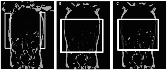
Methods: The children engaged in a series of practice tests to become acclimated to the scanner noises, reduce claustrophobia, and rehearse holding still for a set time. The practice tests assessed if the child could remain still for two minutes while watching a video, first while lying on a blanket, second, on the blanket with headphones, and third, in the mock scanner. The children who passed the three practice tests were then scanned with a 3T Siemens Skyra magnet. Abdominal fat distribution (region of interest (ROI) from the top of the ileac crest to the bottom of the ribcage) volume was measured using 2-point DIXON technique. This region was chosen to give an indication of the body composition around the liver.
Results: Twelve out of eighteen participants successfully completed the actual MRI scan. Chi-squared test showed no significant difference between male and female pass-fail rates. The median age of completed scans was 36 months, whereas the median age for children unable to complete a scan was 28 months. The average total trunk fat was 240.9±85.2mL and the average total VAT was 37.7±25.9mLand liver fat was not quantifiable due to physiological motion. Several strategies (modeling, videos, and incentives) were identified to improve pediatric imaging in different age ranges.
Conclusion: Using an age-specific and tailored protocol, we were able to successfully use MRI for fat imaging in a majority of young children. Development of such protocols enables researchers to better understand the etiology of fat deposition in young children, which can be used to aid in the prevention and treatment of adiposity.
Conflict of interest statement
Figures

Similar articles
-
Measurements of total and regional body composition in preschool children: A comparison of MRI, DXA, and anthropometric data.Obesity (Silver Spring). 2013 May;21(5):1018-24. doi: 10.1002/oby.20205. Obesity (Silver Spring). 2013. PMID: 23784906
-
Relationship of hepatic steatosis to adipose tissue distribution in pediatric nonalcoholic fatty liver disease.J Pediatr Gastroenterol Nutr. 2006 Jan;42(1):83-8. J Pediatr Gastroenterol Nutr. 2006. PMID: 16385259
-
Reproducibility and repeatability of MRI-based body composition analysis.Magn Reson Med. 2020 Dec;84(6):3146-3156. doi: 10.1002/mrm.28360. Epub 2020 Jun 10. Magn Reson Med. 2020. PMID: 32519807
-
Fast, free-breathing and motion-minimized techniques for pediatric body magnetic resonance imaging.Pediatr Radiol. 2018 Aug;48(9):1197-1208. doi: 10.1007/s00247-018-4116-x. Epub 2018 Aug 4. Pediatr Radiol. 2018. PMID: 30078042 Review.
-
Pediatric obesity phenotyping by magnetic resonance methods.Curr Opin Clin Nutr Metab Care. 2005 Nov;8(6):595-601. Curr Opin Clin Nutr Metab Care. 2005. PMID: 16205458 Free PMC article. Review.
Cited by
-
A Protocol for Sedation Free MRI and PET Imaging in Adults with Autism Spectrum Disorder.J Autism Dev Disord. 2019 Jul;49(7):3036-3044. doi: 10.1007/s10803-019-04010-3. J Autism Dev Disord. 2019. PMID: 31004246
-
Imaging of body composition in children.Quant Imaging Med Surg. 2020 Aug;10(8):1661-1671. doi: 10.21037/qims.2020.04.06. Quant Imaging Med Surg. 2020. PMID: 32742959 Free PMC article. Review.
References
-
- Gastaldelli A. Abdominal fat: does it predict the development of type 2 diabetes? Am J Clin Nutr. 2008;87: 1118–1119. Available: http://ajcn.nutrition.org/content/87/5/1118.full - PubMed
Publication types
MeSH terms
LinkOut - more resources
Full Text Sources
Other Literature Sources
Medical
