. 2016 Jun;30(6):2161-70.
doi: 10.1096/fj.201500199R. Epub 2016 Feb 22.

Interplay of the physical microenvironment, contact guidance, and intracellular signaling in cell decision making

Affiliations

Interplay of the physical microenvironment, contact guidance, and intracellular signaling in cell decision making

Colin D Paul et al. FASEB J. 2016 Jun.

Abstract

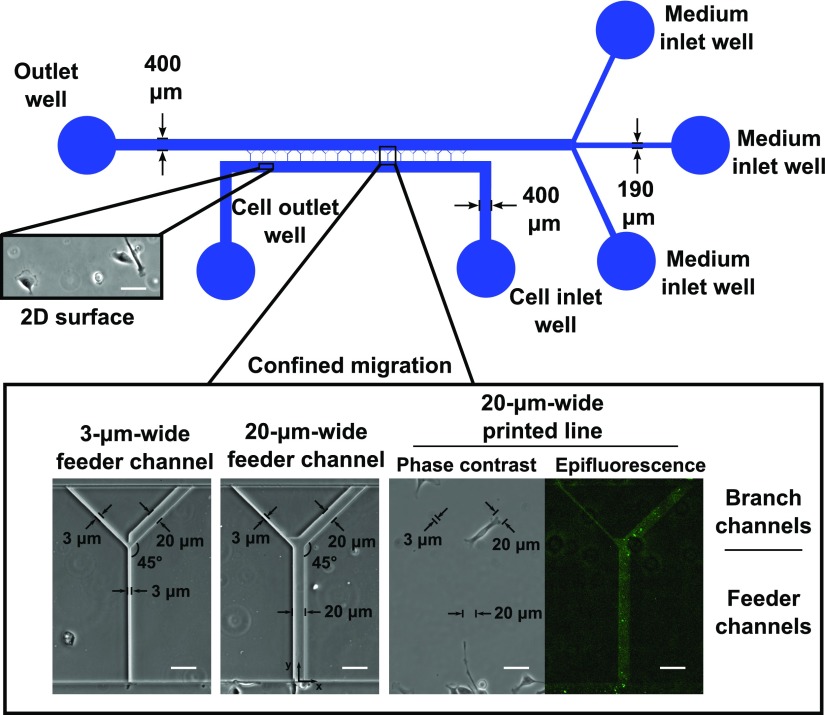
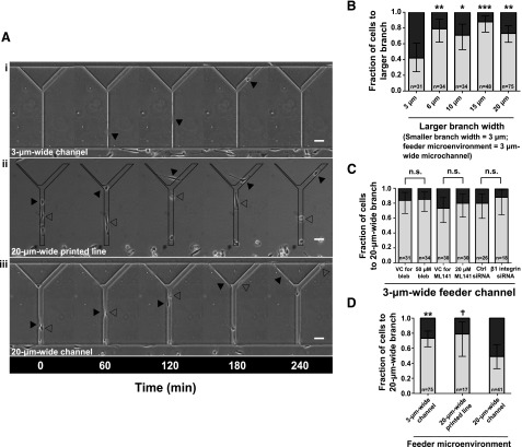
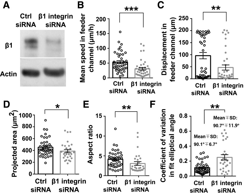
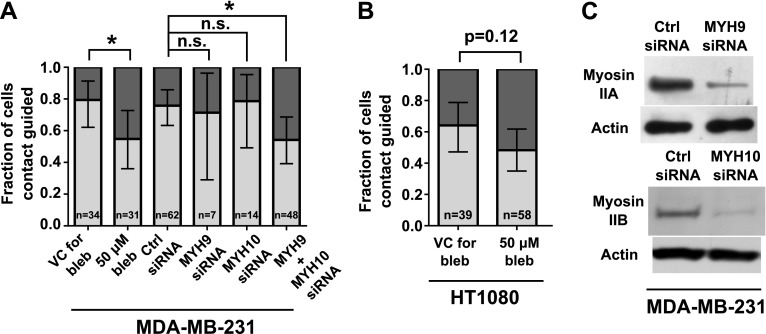
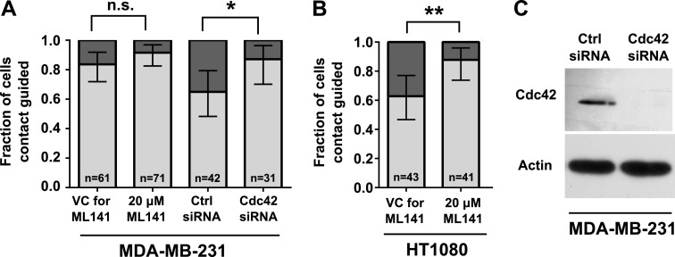
The peritumoral physical microenvironment consists of complex topographies that influence cell migration. Cell decision making, upon encountering anisotropic, physiologically relevant physical cues, has yet to be elucidated. By integrating microfabrication with cell and molecular biology techniques, we provide a quantitative and mechanistic analysis of cell decision making in a variety of well-defined physical microenvironments. We used MDA-MB-231 breast carcinoma and HT1080 fibrosarcoma as cell models. Cell decision making after lateral confinement in 2-dimensional microcontact printed lines is governed by branch width at bifurcations. Cells confined in narrow feeder microchannels prefer to enter wider branches at bifurcations. In contrast, in feeder channels that are wider than the cell body, cells elongate along one side wall of the channel and are guided by contact with the wall to the contiguous branch channel independent of its width. Knockdown of β1-integrins or inhibition of cellular contractility suppresses contact guidance. Concurrent, but not individual, knockdown of nonmuscle myosin isoforms IIA and IIB also decreases contact guidance, which suggests the existence of a compensatory mechanism between myosin IIA and myosin IIB. Conversely, knockdown or inhibition of cell division control protein 42 homolog promotes contact guidance-mediated decision making. Taken together, the dimensionality, length scales of the physical microenvironment, and intrinsic cell signaling regulate cell decision making at intersections.-Paul, C. D., Shea, D. J., Mahoney, M. R., Chai, A., Laney, V., Hung, W.-C., Konstantopoulos, K. Interplay of the physical microenvironment, contact guidance, and intracellular signaling in cell decision making.

Keywords: cell migration; confinement; microfluidics.


Figures

Figure 1.
Figure 1.
Design of microfluidic device and microcontact printed surfaces. To-scale schematic of the microfluidic device used for migration studies and as a template to print collagen type I on glass coverslips. Insets show specific regions of the device. Unconfined 2D surfaces were available in the cell seeding region below the microchannel entrances. Microchannels were arrayed between larger cell seeding and medium channels. Microchannels consisted of 3- or 20-µm-wide feeder channels bifurcating to branch channels of various widths. All microchannels were 10 µm in height. Alternatively, collagen type I was printed in the same projected geometry as that of the microchannels. Deposited collagen is shown in the epifluorescence image. Scale bars, 50 µm.
Figure 2.
Figure 2.
Cell decision making from various feeder microenvironments. The physical microenvironment affects the decision making of MDA-MB-231 cells at bifurcations. A) Phase contrast images of MDA-MB-231 cells migrating inside Y-shaped microchannels with 3-µm-wide feeder channels bifurcating to 3- and 20-µm-wide branches (i); on Y-shaped printed lines with a 20-µm-wide feeder region bifurcating to 3- and 20-µm-wide branches (ii); and inside Y-shaped microchannels with 20-µm-wide feeder channels bifurcating to 3- and 20-µm-wide branches (iii). All surfaces were coated with collagen type I (20 µg/ml). Arrowheads illustrate the positions of indicated cells at prescribed time points. B) Fraction of cells from a 3-µm-wide feeder microchannel migrating into the larger branch when encountering a bifurcation in which the width of 1 branch was set at 3 µm and the width of the other branch varied from 3 to 6, 10, 15, or 20 µm. C) Fraction of cells entering the 20-µm-wide branch channels from a 3-µm-wide feeder channel upon treatment with 50 µM blebbistatin, 20 µM ML141, or the appropriate controls, or transfection with nontargeting control (ctrl) siRNA or siRNA targeting β1-integrin. D) Fraction of cells from the microenvironments shown in panel A entering the 20-µm-wide branch at bifurcations. In panels B-D, data represent fraction of cells. Error bars show 95% confidence intervals of these fractions. The total number of cells making a decision for each condition is indicated on the plot. Data were collected and pooled over n ≥ 4 independent experiments. Significance was assessed by using the 2-population proportion z test with respect to the design containing symmetric 3-µm-wide branch channels (B), the device with the 20-µm-wide feeder microchannel (C), or to control cells (D). n.s., not significant. P < 0.1; *P < 0.05; **P < 0.01; ***P < 0.001. Scale bars, 50 µm.
Figure 3.
Figure 3.
The distinct role of contact guidance in cell decision making at bifurcations and polarization in microchannels. A) Schematic of cell paths for a contact-guided and a noncontact-guided cell. Cells were scored as contact guided if they did not cross the midline of the channel while in the bifurcation region of the device. The bifurcation region (box) is defined as the area within 1 cell length away from the bifurcation. B) Fraction of contact-guided MDA-MB-231 and HT1080 cells in 20-µm-wide feeder microchannels. In select experiments with MDA-MB-231 cells, the glass slide making up the base of the microchannels was coated with a thin layer (∼25 µm) of PDMS before device assembly and functionalization with collagen type I. Columns show fraction of contact-guided cells, and bars show 95% confidence intervals of the fractions. Comparison between proportions of contact-guided MDA-MB-231 cells migrating on glass or PDMS bases was made using the 2-population proportion z test. CF) Circularity (C), average speed (D) elliptical angle of fit (E), and minor axis length (F) of MDA-MB-231 cells that go on to enter a branch channel as a function of distance into the feeder channel. Measurements were binned within 20-µm-long regions of the feeder channel. Symbols show the average value in each bin, accompanied by se of each measurement. Symbols are plotted at the center position of each bin. Dashed line (E) shows the angle of the microchannel (90° with respect to the horizontal). All data were collected and pooled over n ≥ 2 independent experiments. G) Representative images of MDA-MB-231 cells migrating in 20-µm-wide, 10-µm-tall feeder microchannels. Cells were stained for F-actin using Alexa Fluor 488 phalloidin. Confocal images taken at the basal surface of the cell are shown in the x-y plane. Cells were imaged at 0.8 µm axial intervals to generate orthogonal reconstructions in the x-z and y-z planes. The locations of the orthogonal reconstructions are indicated by the white lines in the x-y planes. A 3D surface reconstruction of the indicated cell is shown in the inset. n.s., not significant. Scale bars, 50 µm (A) and 20 µm (G).
Figure 4.
Figure 4.
The critical role of integrin-mediated adhesions in the contact guidance of MDA-MB-231 cells. MDA-MB-231 cells were transfected with nontargeting control (ctrl) siRNA or siRNA-targeting β1-integrin. A) β1-integrin knockdown was confirmed via Western blot. BF) Average speed (B), displacement (C), projected area (D), aspect ratio (E), and coefficient of variation in elliptical angle of fit (F) of cells transfected with ctrl or β1-integrin siRNA. Parameters were quantified in the straight feeder regions of the microchannels. Columns represent population means. Error bars show sem. Data points represent values of each metric for 1 cell for n ≥ 28 cells from n = 4 independent experiments. *P < 0.05; **P < 0.01; ***P < 0.001 by Mann-Whitney U test.
Figure 5.
Figure 5.
Inhibition of cell contractility impairs contact guidance–mediated decision making of MDA-MB-231 and HT1080 cells. MDA-MB-231 or HT1080 cells were treated with 50 µM blebbistatin or VC. In select experiments, MDA-MB-231 cells were transfected with control (ctrl) siRNA or siRNA-targeting myosin IIA (MYH9) and/or myosin IIB (MYH10). A, B) Fractions of contact-guided MDA-MB-231 cells (A) and HT1080 cells (B) during the decision-making process from 20-µm-wide feeder channels. Data show overall fraction of contact-guided cells, and bars show 95% confidence intervals of the fractions. Data were collected and pooled over n ≥ 3 independent experiments. The number of cells assayed for each condition is indicated on the plot. Comparisons between fractions of treated and control cells were analyzed using the 2-population proportion z test. C) Knockdown of myosin isoforms in MDA-MB-231 cells was confirmed via Western blot, with actin as loading control. Panel shows 2 blots from the same cell lysate (collected after concurrent knockdown of myosin IIA and myosin IIB) immunoblotted by using an antibody to either myosin IIA (MYH9) or myosin IIB (MYH10). n.s., not significant. *P < 0.05.
Figure 6.
Figure 6.
Inhibition of Cdc42 increases contact guidance–mediated decision making of MDA-MB-231 and HT1080 cells. MDA-MB-231 or HT1080 cells were treated with ML141 (20 µM) or VC. In separate experiments, MDA-MB-231 cells were transfected with control (ctrl) siRNA or siRNA-targeting Cdc42. A, B) Fractions of contact-guided MDA-MB-231 cells (A) and HT1080 cells (B) during the decision-making process. Data show overall fraction of contact-guided cells, and bars show 95% confidence intervals of the fractions. Data were collected and pooled over n ≥ 3 independent experiments. The number of cells assayed for each condition is indicated on the plot. Comparisons between fractions of treated and control cells were analyzed using the 2-population proportion z test. C) Knockdown of Cdc42 in MDA-MB-231 cells was confirmed via Western blot, with actin as loading control. n.s., not significant. *P < 0.05.

References

    1. Alexander S., Koehl G. E., Hirschberg M., Geissler E. K., Friedl P. (2008) Dynamic imaging of cancer growth and invasion: a modified skin-fold chamber model. Histochem. Cell Biol. 130, 1147–1154 - PubMed
    1. Weigelin B., Bakker G.-J., Friedl P. (2012) Intravital third harmonic generation microscopy of collective melanoma cell invasion: principles of interface guidance and microvesicle dynamics. Intravital 1, 32–43 - PMC - PubMed
    1. Stroka K. M., Konstantopoulos K. (2014) Physical biology in cancer. 4. Physical cues guide tumor cell adhesion and migration. Am. J. Physiol. Cell Physiol. 306, C98–C109 - PMC - PubMed
    1. Alexander S., Weigelin B., Winkler F., Friedl P. (2013) Preclinical intravital microscopy of the tumour-stroma interface: invasion, metastasis, and therapy response. Curr. Opin. Cell Biol. 25, 659–671 - PubMed
    1. Wolf K., Alexander S., Schacht V., Coussens L. M., von Andrian U. H., van Rheenen J., Deryugina E., Friedl P. (2009) Collagen-based cell migration models in vitro and in vivo. Semin. Cell Dev. Biol. 20, 931–941 - PMC - PubMed

Publication types