. 2016 Feb 23:7:10832.
doi: 10.1038/ncomms10832.

Vector-free transmission and persistence of Japanese encephalitis virus in pigs

Affiliations

Vector-free transmission and persistence of Japanese encephalitis virus in pigs

Meret E Ricklin et al. Nat Commun. .

Abstract

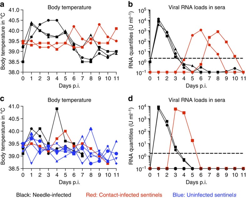
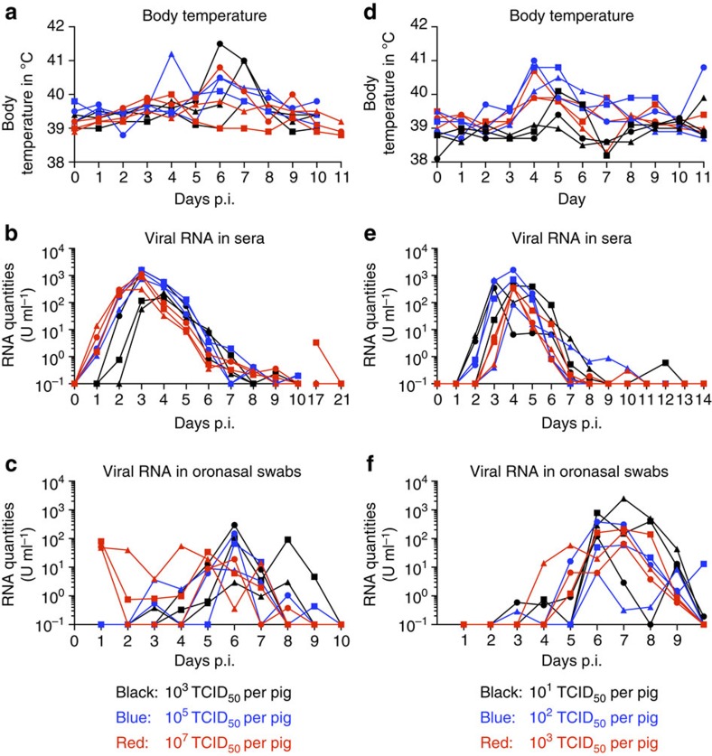
Japanese encephalitis virus (JEV), a main cause of severe viral encephalitis in humans, has a complex ecology, composed of a cycle involving primarily waterbirds and mosquitoes, as well as a cycle involving pigs as amplifying hosts. To date, JEV transmission has been exclusively described as being mosquito-mediated. Here we demonstrate that JEV can be transmitted between pigs in the absence of arthropod vectors. Pigs shed virus in oronasal secretions and are highly susceptible to oronasal infection. Clinical symptoms, virus tropism and central nervous system histological lesions are similar in pigs infected through needle, contact or oronasal inoculation. In all cases, a particularly important site of replication are the tonsils, in which JEV is found to persist for at least 25 days despite the presence of high levels of neutralizing antibodies. Our findings could have a major impact on the ecology of JEV in temperate regions with short mosquito seasons.


Figures

Figure 1
Figure 1. Body temperature and viraemia of pigs infected by needle or contact.
(a,b) Three needle-infected pigs (107 TCID50 per pig, black lines) were housed with two naive sentinel animals (red lines). (c,d) Two animals were needle-infected (black) and housed with six sentinel pigs (red and blue). Viraemia is shown as viral RNA loads determined by real-time RT–PCR, and expressed as U ml−1 (1 U corresponding to the RNA quantity found in 1 TCID50 of a virus stock).
Figure 2
Figure 2. Viral RNA loads in peripheral and CNS tissues.
Viral RNA loads in peripheral (a) and CNS (b) tissues were determined at necropsy by real-time RT–PCR and expressed as U g−1 (1 U corresponding to the RNA quantity found in 1 TCID50 of a virus stock). Solid symbols represent needle-infected pigs (n=5) killed at 7 (circles) and 11 (squares) days p.i. Open symbols represent pigs infected by contact (n=3). Two animals, corresponding to those shown in Fig. 1b, were killed at day 11, which was 4 and 7 days after the peak of viraemia, respectively. One animal, corresponding to Fig. 1d, was killed at day 10, which was 7 days after peak viraemia. Asterisks (*) indicate significant differences calculated with a nonparametric two-tailed Mann–Whitney U-test (P<0.05).
Figure 3
Figure 3. Viral RNA load in oronasal swabs.
Oronasal swabs were collected daily from the experiments shown in Fig. 1 (experiment one and two as described in Methods); viral loads were quantified by real-time RT–qPCR (1 U corresponding to the RNA quantity found in 1 TCID50 of a virus stock). Black: needle-infected animals (n=5); red: contact-infected pigs (n=3).
Figure 4
Figure 4. Body temperature and viral RNA loads in serum and swabs of oronasally infected pigs.
In ac, groups of three animals were infected oronasally with JEV at 103 (black), 105 (blue) or 107 (red) TCID50 per pig. (df) A second experiment with groups of three animals infected oronasally at 101 (black), 102 (blue) or 103 (red) TCID50 per pig. Viral RNA loads in serum (d,e) and oronasal swabs (c,f) were determined as for the other figures, and are expressed as U ml−1.
Figure 5
Figure 5. Virus tropism in oronasally infected pigs.
Viral load in peripheral organs (a) and CNS (b) of oronasally infected pigs was assessed at 10 days p.i. by real-time RT–PCR, and shown in relative RNA quantities (U ml−1). Animals were infected with either 103 (filled circles) or 105 (open circles) TCID50. Asterisks (*) indicate significant differences calculated with a nonparametric two-tailed Mann–Whitney U-test (P<0.05).
Figure 6
Figure 6. CNS lesions in pigs are not dependent on the route of infection.
Lesions in the central nervous system were semiquantitatively scored from 0 to 4 (0: no lesions; 1: minimal lesions; 2: mild lesions; 3: moderate lesions; 4: severe lesions) as described in the Methods section. The needle-infected group (needle) was analysed 6 and 10 days after the onset of viraemia (taken as a measure due to the unknown infection time and different incubation periods of the other groups). Tissue from pigs infected by contact (contact) was 4, 6 and 7 days after the onset of viraemia. Tissue from oronasally infected animals was 7, 8 and 9 days after the onset of viraemia. CNS tissue from the animals infected with 103 TCID50 (oronasal 10E3) and 105 TCID50 (oronasal 10E5) is shown. olf., olfactory.
Figure 7
Figure 7. Persistence of JEV in the tonsils.
(a) Various tissues were collected from oronasally infected pigs (107 TCID50, Nakayama strain) and analysed for viral RNA load by real-time RT–PCR. The animal depicted by red squares is identical to the animal depicted in Fig. 4b, which became viraemic at day 17 p.i. but cleared virus from the blood by day 21 p.i. (b,c). Six pigs were needle-infected with the Laos strain of JEV, and tonsils were analysed for viral RNA load at 11 and 25 days p.i. Viral RNA loads were determined as for the other figures and expressed as U ml−1. Asterisks (*) indicate significant differences calculated with a nonparametric two-tailed Mann–Whitney U-test (P<0.05). Neoc., neocortex.
Figure 8
Figure 8. Virus neutralization titres.
Sera obtained from experimentally infected groups were obtained at the indicated days (d) p.i. and tested in a PRNT test. Needle: animals infected with the Nakayama strain (Fig. 1); contact: animals infected by contact (see Fig. 1, time point p.i. is estimated); ON: pigs infected via the oronasal route (Fig. 4); Laos: pigs infected i.d. with the Laos strain of JEV (106 TCID50).

References

    1. Solomon T. et al. Japanese encephalitis. J. Neurol. Neurosurg. Psychiatry 68, 405–415 (2000). - PMC - PubMed
    1. Impoinvil D. E., Baylis M. & Solomon T. Japanese encephalitis: on the One Health agenda. Curr. Top. Microbiol. Immunol. 365, 205–247 (2013). - PubMed
    1. van den Hurk A. F., Ritchie S. A. & Mackenzie J. S. Ecology and geographical expansion of Japanese encephalitis virus. Annu. Rev. Entomol. 54, 17–35 (2009). - PubMed
    1. Campbell G. L. et al. Estimated global incidence of Japanese encephalitis: a systematic review. Bull. World Health Organ. 89, 766–774 774a–774e (2011). - PMC - PubMed
    1. Weaver S. C. & Barrett A. D. Transmission cycles, host range, evolution and emergence of arboviral disease. Nat. Rev. Microbiol. 2, 789–801 (2004). - PMC - PubMed

Publication types