. 2016 Feb 23:6:21949.
doi: 10.1038/srep21949.

The Structural Basis of Oncogenic Mutations G12, G13 and Q61 in Small GTPase K-Ras4B

Affiliations

The Structural Basis of Oncogenic Mutations G12, G13 and Q61 in Small GTPase K-Ras4B

Shaoyong Lu et al. Sci Rep. .

Abstract

Ras mediates cell proliferation, survival and differentiation. Mutations in K-Ras4B are predominant at residues G12, G13 and Q61. Even though all impair GAP-assisted GTP → GDP hydrolysis, the mutation frequencies of K-Ras4B in human cancers vary. Here we aim to figure out their mechanisms and differential oncogenicity. In total, we performed 6.4 μs molecular dynamics simulations on the wild-type K-Ras4B (K-Ras4B(WT)-GTP/GDP) catalytic domain, the K-Ras4B(WT)-GTP-GAP complex, and the mutants (K-Ras4B(G12C/G12D/G12V)-GTP/GDP, K-Ras4B(G13D)-GTP/GDP, K-Ras4B(Q61H)-GTP/GDP) and their complexes with GAP. In addition, we simulated 'exchanged' nucleotide states. These comprehensive simulations reveal that in solution K-Ras4B(WT)-GTP exists in two, active and inactive, conformations. Oncogenic mutations differentially elicit an inactive-to-active conformational transition in K-Ras4B-GTP; in K-Ras4B(G12C/G12D)-GDP they expose the bound nucleotide which facilitates the GDP-to-GTP exchange. These mechanisms may help elucidate the differential mutational statistics in K-Ras4B-driven cancers. Exchanged nucleotide simulations reveal that the conformational transition is more accessible in the GTP-to-GDP than in the GDP-to-GTP exchange. Importantly, GAP not only donates its R789 arginine finger, but stabilizes the catalytically-competent conformation and pre-organizes catalytic residue Q61; mutations disturb the R789/Q61 organization, impairing GAP-mediated GTP hydrolysis. Together, our simulations help provide a mechanistic explanation of key mutational events in one of the most oncogenic proteins in cancer.


Figures

Figure 1
Figure 1. The architecture of GppNHp- and GDP-bound K-Ras4B catalytic domain.
(A) The Kabsch-Sander secondary structure cartoon of the K-Ras4B catalytic domain. The blue solid arrows represent β-strands, the red solid cylinders represent α-helices and the gray solid cylinders represent loops. (B) Cartoon representation of crystal structure of GppNHp-bound K-Ras4B (PDB ID: 2PMX). The helices, strands and loops are colored by red, blue and gray, respectively. (C) Arrangements of active site residues G13, Y32 and Q61 in the GppNHp-bound K-Ras4B (D) Surface representation of GppNHp-bound K-Ras4B. (E) Arrangements of residues G13, Y32 and Q61 in the GDP-bound K-Ras4B (PDB ID: 4LPK). (F) Surface representation of GDP-bound K-Ras4B. (G) Backbone superimposition of GppNHp- (magenta) and GDP-bound (cyan) K-Ras4B. The P-loop, switch I and switch II domains, are colored by green, pink and light blue, respectively. Mg2+ ion is depicted by a green sphere.
Figure 2
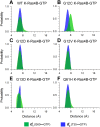
Figure 2. Oncogenic mutations shift the population of K-Ras4B–GTP from the inactive to the active state.
The probability distributions for two atom-pairs distances, d1 (defined by the distance from G60 Cα atom to GTP Pβ atom) and d2 (defined by the distance from T35 Cα atom to GTP Pβ atom), were calculated on the MD snapshots of K-Ras-GTP. (A) wild-type, (B) G12C, (C) G12D, (D) G12V, (E) G13D and (F) Q61H mutants.
Figure 3
Figure 3. G12C and G12D mutations significantly affect the conformational ensemble of K-Ras4B-GDP.
The probability distributions for two atom-pairs distances, d1 (defined by the distance from G60 Cα atom to GDP Pβ atom) and d2 (defined by the distance from T35 Cα atom to GDP Pβ atom), were calculated on the MD snapshots of K-Ras4B-GDP. (A) wild-type, (B) G12C, (C) G12D, (D) G12V, (E) G13D and (F) Q61H mutants. K-Ras4BWT-GDP exhibits one major energy-minima basin, corresponding to the inactive state. Oncogenic G12 mutations, particularly G12C and G12D, result in larger conformational changes of K-Ras4B-GDP.
Figure 4
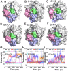
Figure 4. Oncogenic mutations affect the dynamics of GDP-binding site.
Surface representation of the representative structures of the wild-type (A), G12C (B), G12D (C), G12V (D), G13D (E) and Q61H (F) mutants. Time dependence of interresidue distances between the Cα atoms of G12/P34 (G), G12/G60 (H), and G13/E61 (I) residue pairs. The oncogenic G12C and G12D mutations cause larger exposure of the nucleotide-binding site compared to the wild-type and other mutations.
Figure 5
Figure 5. G12C and G12D mutations increase the exposure of the GDP-binding site.
The solvent accessible surface area (SASA, Å2) of GDP in the wild-type and oncogenic mutants.
Figure 6
Figure 6. The conformational transition of K-Ras4B is more accessible in the GTP-to-GDP exchange than in the GDP-to-GTP exchange.
(A) Time dependence of distances between T35 OG1 atom and GDP O1B atom as well as between G60 N atom and GDP O3B atom in both the K-Ras4BWT-GTP simulation (0–400 ns) and the GTP-to-GDP exchange simulation (400–600 ns). Surface representation of the representative structures of K-Ras4B derived from 400–550 ns (B) and 550–600 ns (C). (D) The SASA (Å2) of GDP in the periods of 400–550 ns and 550–600 ns. (E) Time dependence of distances between T35 OG1 atom and GTP O2G atom as well as between G60 N atom and GTP O1G atom in both the K-Ras4BWT-GDP simulation (0–400 ns) and the GDP-to-GTP exchange simulation (400–600 ns). (F) Surface representation of the representative structure in the period of 400–600 ns.
Figure 7
Figure 7. GAP binding shifts the conformational ensemble of K-Ras4BWT-GTP to the GTP-bound active state.
The probability distributions for two atom-pairs distances, d1 and d2, were calculated on the MD snapshots of K-Ras4BWT-GTP–GAP.
Figure 8
Figure 8. GAP stabilizes the active, catalytically competent conformation of K-Ras4BWT–GTP.
(A) Details of interactions of K-Ras4B with GAP highlighting catalytically important elements. In the K-Ras4BWT–GTP complex, GAP provides the arginine finger R789 to interact with the α- and γ-phosphates of GTP. Meanwhile, the side chain carbonyl group of catalytic residue Q61 interacts with the catalytic water. In this conformer, Q61 can extract a hydrogen atom from the catalytic water, and the developing negative hydroxyl ion can attack the γ-phosphorus of GTP resulting in GAP-mediated GTP hydrolysis. (B) The evolution of the catalytic water molecule (WAT) to Q61 and GTP in the active site.
Figure 9
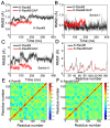
Figure 9. GAP stabilizes the catalytically-competent conformation of K-Ras4B in addition to providing the arginine finger R789 for catalysis.
(A) The Cα atoms RMSD of K-Ras4B in the free K-Ras4B and K-Ras4B–GAP complex. The Cα atoms RMSD of K-Ras4B switch I (B) and switch II (C) regions in the free K-Ras4B and K-Ras4B–GAP complex. (D) The Cα atoms RMSF of K-Ras4B in the free K-Ras4B and K-Ras4B–GAP complex. The extent of correlation for all residue pairs (of Cα atom displacement) of K-Ras4B in the free K-Ras4B (E) and K-Ras4B–GAP complex (F). The domain-domain motions are markedly restricted in the K-Ras4B–GAP complex compared to the free K-Ras4B, particularly in the switch II domain (residues 59–67).
Figure 10
Figure 10. Oncogenic mutations disturb the catalytically-competent arrangements of Q61 and R789.
Structural view of the active site from the representative structures of G12C (A), G12D (B), G12V (C), G13D (D) and Q61H (E) K-Ras4B-GTPGAP complex. The salt bridge and H-bonding interactions are depicted by blue and wheat dotted lines, respectively. (F) Time dependence of the distance between Q61 OE1 atom and GTP Pγ atom (in Q61H mutant, the distance was measured between H61 ND1 atom and GTP Pγ atom) in the wild-type and oncogenic mutants. The G12C mutation significantly disturbs the arrangement of R789 where it moves away from GTP and of Q61, where it cannot form H-bond with the γ-phosphate. The G12D and G12V mutations cause rearrangement of the side chain OE1 atom of Q61 where it cannot extract a hydrogen atom from the catalytic water molecule. The G13D mutation abolishes the direct interactions between the side chain of Q61 and GTP. The Q61H mutation increases the distance between the side chain ND1 atom of H61 and γ-phosphorus and changes the angle among the atoms NE2 and ND1 of Q61 and γ-phosphorus, leading to the inability of Q61 to coordinate the catalytic water molecule.

References

    1. Milburn M. V. et al.. Molecular switch for signal transduction: structural differences between active and inactive forms of protooncogenic ras proteins. Science 247, 939–45 (1990). - PubMed
    1. Cherfils J. & Zeghouf M. Regulation of small GTPases by GEFs, GAPs, and GDIs. Physiol. Rev. 93, 269–309 (2013). - PubMed
    1. Bos J. L., Rehmann H. & Wittinghofer A. Review GEFs and GAPs: Critical elements in the control of small G proteins. Cell 129, 865–877 (2007). - PubMed
    1. Sondermann H. et al.. Structural Analysis of Autoinhibition in the Ras Activator Son of Sevenless. Cell 119, 393–405 (2004). - PubMed
    1. Jang H. et al.. The higher level of complexity of K-Ras4B activation at the membrane. FASEB J. doi: 10.1096/fj.15–279091 (2015). - DOI - PMC - PubMed

Publication types

MeSH terms