. 2016 Feb 22;5(2):e002767.
doi: 10.1161/JAHA.115.002767.

Trimethylamine N-Oxide Promotes Vascular Inflammation Through Signaling of Mitogen-Activated Protein Kinase and Nuclear Factor-κB

Affiliations

Trimethylamine N-Oxide Promotes Vascular Inflammation Through Signaling of Mitogen-Activated Protein Kinase and Nuclear Factor-κB

Marcus M Seldin et al. J Am Heart Assoc. .

Abstract

Background: The choline-derived metabolite trimethylamine N-oxide (TMAO) has been demonstrated to contribute to atherosclerosis and is associated with coronary artery disease risk.

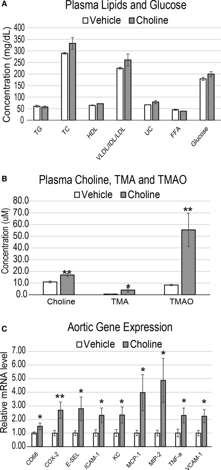
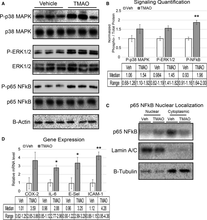
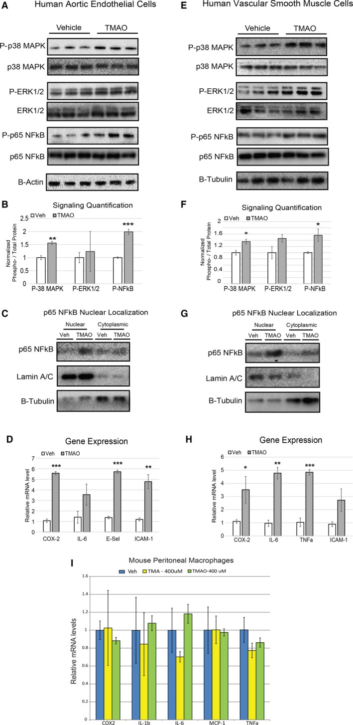
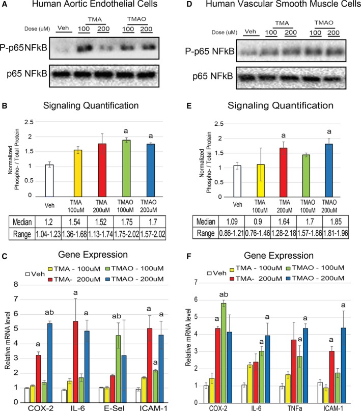
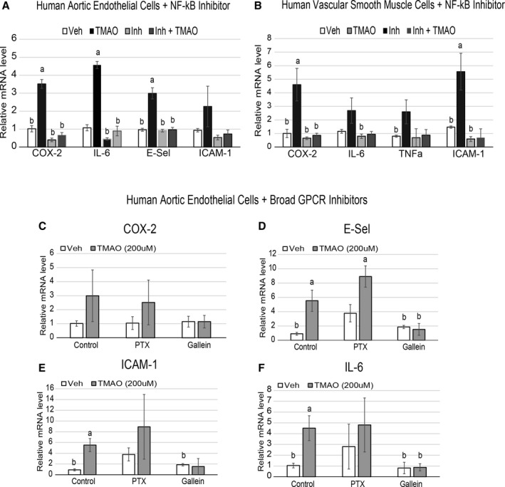
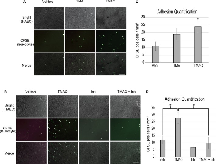
Methods and results: We explored the impact of TMAO on endothelial and smooth muscle cell function in vivo, focusing on disease-relevant outcomes for atherogenesis. Initially, we observed that aortas of LDLR(-/-) mice fed a choline diet showed elevated inflammatory gene expression compared with controls. Acute TMAO injection at physiological levels was sufficient to induce the same inflammatory markers and activate the well-known mitogen-activated protein kinase, extracellular signal-related kinase, and nuclear factor-κB signaling cascade. These observations were recapitulated in primary human aortic endothelial cells and vascular smooth muscle cells. We also found that TMAO promotes recruitment of activated leukocytes to endothelial cells. Through pharmacological inhibition, we further showed that activation of nuclear factor-κB signaling was necessary for TMAO to induce inflammatory gene expression in both of these relevant cell types as well as endothelial cell adhesion of leukocytes.

Conclusions: Our results suggest a likely contributory mechanism for TMAO-dependent enhancement in atherosclerosis and cardiovascular risks.

Keywords: atherosclerosis; cardiovascular disease; endothelial cell; inflammation; leukocyte adhesion; nuclear factor‐κB signaling; trimethylamine N‐oxide; vascular smooth muscle cell.


Figures

Figure 1
Figure 1
Choline feeding causes inflammation in atherosclerosis‐prone mouse aortas that is accompanied by increased plasma choline, TMA, and TMAO but not lipids. A through C, LDLR −/− female mice were fed chow or chow with either 0.07% (vehicle) or 1.3% choline for 3 weeks. Plasma was quantified for circulating levels of TG, TC, HDL, VLDL/IDL/LDL, UC, FFA, and glucose (A), as well as choline, TMA, and TMAO (B). Then aortas were harvested and quantitative polymerase chain reaction was used to quantify expression levels of inflammatory genes (C). All genes expressed as mean±SEM and normalized to RPL13A expression. n=8 mice per group; *P<0.05; **P<0.01. COX‐2 indicates cyclooxygenase 2; E‐SEL, E‐selectin; FFA, free fatty acids; HDL, high‐density lipoprotein; IDL, xxx; LDL, low‐density lipoprotein; MCP‐1, monocyte chemotactic protein 1; MIP‐2, macrophage inflammatory protein 2; TC, total cholesterol; TG, triglyceride; TMA, trimethylamine; TMAO, trimethylamine N‐oxide; TNF‐a, tumor necrosis factor α; UC, unesterified cholesterol; VLDL, very low‐density lipoprotein.
Figure 2
Figure 2
Acute TMAO injection causes activation of signaling cascades and elevated inflammatory gene expression. A and B, LDLR −/− female mice were fasted for 4 hours and then injected with vehicle or TMAO (86 μmol). Aortas were harvested 30 minutes following injection and immunoblotted (A) and quantified (B) for phosphorylation of p38 MAPK (Thr‐180/Tyr‐182), ERK1/2 (Thr‐202/Try‐204), and p65 NFkB (Ser536). n=3, nonparametric t test using a Mann–Whitney U test was performed for comparisons. Median and range are reported for each group. C, Lysate from the same aortas was fractionated into nuclear and cytosolic compartments and immune‐probed for the presence of p65 NFkB with lamin A/C and β‐tubulin, indicating the purity of the nuclear and cytosolic fractions, respectively. D, A separate cohort of LDLR −/− mice underwent the same fasting–injection procedure, and then aortas were harvested 5 hours after injection, and qPCR was used to quantify expression levels of inflammatory genes. All data were expressed as mean±SEM. Immunoblot quantification was normalized to P‐residue/total protein, with β‐actin serving as a loading control; qPCR genes normalized to RPL13A expression. n=3, nonparametric t test using a Mann–Whitney U test was performed for comparisons. Median and range are reported for each group; *P<0.05; **P<0.01. ERK1/2 indicates extracellular signal–related kinase 1/2; MAPK, mitogen‐activated protein kinase; NFkB, nuclear factor‐κB; qPCR, quantitative polymerase chain reaction; TMAO, trimethylamine N‐oxide; TNF‐a, tumor necrosis factor α; Veh, vehicle.
Figure 3
Figure 3
TMAO induces inflammation cascades and transcription in primary human endothelial and smooth muscle cells. A through C, Immunoblots (A), corresponding quantifications (B), and cell fractionations (C) of HAECs treated with vehicle (PBS) or TMAO (100 μmol/L) for 40 minutes (n=6). D, HAECs were treated with vehicle (PBS) or TMAO (200 μmol/L) for 6 hours and then quantified for inflammatory gene expression (n=6). E through H, The same experimental conditions were used as in panels A through D with primary VSMCs instead of HAECs. All data are expressed as mean±SEM. Immunoblot quantification was normalized to P‐residue/total protein. β‐actin and β‐tubulin were used as the loading controls for HAECs and VSMCs, respectively, and quantitative polymerase chain reaction genes normalized to RPL13A expression. I, Mouse peritoneal macrophages were treated with indicated amounts of vehicle, trimethylamine, or TMAO (n=4 for each group) and then evaluated for inflammatory gene expression. All experiments were confirmed in at least 3 separate donors to confirm robust function. *P<0.05; **P<0.01; ***P<0.001. COX‐2 indicates cyclooxygenase 2; E‐Sel, E‐selectin; ERK1/2 indicates extracellular signal–related kinase 1/2; HAEC, human aortic endothelial cell; IL‐6, interleukin 6; MAPK, mitogen‐activated protein kinase; NFkB, nuclear factor‐κB; qPCR, quantitative polymerase chain reaction; TMAO, trimethylamine N‐oxide; Veh, vehicle; VSMC, vascular smooth muscle cell.
Figure 4
Figure 4
TMAO is more efficacious than TMA at affecting cell inflammation at physiological concentrations. A and B, HAECs were treated for 40 minutes with indicated concentrations of vehicle, TMA or TMAO, and then immunoblotted (A) and quantified (B) for phosphorylation of p65 NF‐κB (Ser536) (n=3). C, HAECs were treated for 6 hours in the same conditions as were used in panels A and B and then subjected to qPCR for inflammatory gene expression (n=5). D‐F, The same procedures were carried out as were used in panels A through C except with vascular smooth muscle cells instead of HAECs. All experiments were confirmed in at least 3 separate donors, and qPCR genes normalized to RPL13A expression. a P<0.05 vs vehicle treatment group; b P<0.05 of TMAO vs matched concentration of TMA. COX‐2 indicates cyclooxygenase 2; E‐Sel, E‐selectin; HAEC, human aortic endothelial cell; IL‐6, interleukin 6; NFkB, nuclear factor‐κB; qPCR, quantitative polymerase chain reaction; TMA, trimethylamine; TMAO, trimethylamine N‐oxide; TNF‐a, tumor necrosis factor α; Veh, vehicle.
Figure 5
Figure 5
NFkB and Gßγ signaling is required for TMAO‐induced inflammatory gene expression in endothelial and smooth muscle cells. A and B, HAECs (A) or vascular smooth muscle cells (B) were pretreated for 30 minutes with control (DMSO) or 100 nmol/L NFkB activation inhibitor followed by an overnight treatment with or without 200 μmol/L TMAO and probed by qPCR for inflammatory gene expression (n=6). ANOVA with a Tukey post hoc test was used with TMAO and inhibitor as the 2 factors. We did not observe interaction between inhibitor and TMAO groups. C through F, HAECs were pretreated for 1 hour with control (DMSO), 200 ng/mL PTX, or 10 μmol/L Gallein, followed by addition of vehicle (PBS) or 200 μmol/L TMAO, then probed by qPCR for inflammatory gene expression. All experiments were confirmed in at least 3 separate donors, and qPCR genes normalized to RPL13A expression. a P<0.05 compared with vehicle‐alone group; b P<0.05 compared with TMAO‐alone treatment group. COX‐2 indicates cyclooxygenase 2; E‐Sel, E‐selectin; HAEC, human aortic endothelial cell; IL‐6, interleukin 6; NFkB, nuclear factor‐κB; PTX, pertussis toxin; qPCR, quantitative polymerase chain reaction; TMAO, trimethylamine N‐oxide; TNF‐a, tumor necrosis factor α; Veh, vehicle.
Figure 6
Figure 6
TMAO enhances HAEC recruitment of activated leukocytes through NFkB. A, HAECs were stimulated for adhesive processes for 5 hours with vehicle (PBS) or 400 μmol/L TMA or TMAO then presented with prelabeled, activated leukocytes and quantified (C). B, The same procedure as was used in panel A was followed except a 1‐hour inhibitor treatment (NFkB activation inhibitor) step was added prior to stimulation. D, Quantification of adhered leukocytes of panel B. ANOVA with a Tukey post hoc test was used with TMAO and inhibitor as the 2 factors. We did not observe interaction between inhibitor and TMAO groups. Images reflect representative examples of 9 images taken per group using 3 separate HAEC donor samples. Scale bars=100 μm. HAEC indicates human aortic endothelial cell; Inh, inhibitor; NFkB, nuclear factor‐κB; TMA, trimethylamine; TMAO, trimethylamine N‐oxide.

References

    1. Wang Z, Klipfell E, Bennett BJ, Koeth R, Levison BS, Dugar B, Feldstein AE, Britt EB, Fu X, Chung YM, Wu Y, Schauer P, Smith JD, Allayee H, Tang WH, DiDonato JA, Lusis AJ, Hazen SL. Gut flora metabolism of phosphatidylcholine promotes cardiovascular disease. Nature. 2011;472:57–63. - PMC - PubMed
    1. Koeth RA, Wang Z, Levison BS, Buffa JA, Org E, Sheehy BT, Britt EB, Fu X, Wu Y, Li L, Smith JD, DiDonato JA, Chen J, Li H, Wu GD, Lewis JD, Warrier M, Brown JM, Krauss RM, Tang WH, Bushman FD, Lusis AJ, Hazen SL. Intestinal microbiota metabolism of L‐carnitine, a nutrient in red meat, promotes atherosclerosis. Nat Med. 2013;19:576–585. - PMC - PubMed
    1. Tang WH, Wang Z, Levison BS, Koeth RA, Britt EB, Fu X, Wu Y, Hazen SL. Intestinal microbial metabolism of phosphatidylcholine and cardiovascular risk. N Engl J Med. 2013;368:1575–1584. - PMC - PubMed
    1. Wang Z, Tang WH, Buffa JA, Fu X, Britt EB, Koeth RA, Levison BS, Fan Y, Wu Y, Hazen SL. Prognostic value of choline and betaine depends on intestinal microbiota‐generated metabolite trimethylamine‐N‐oxide. Eur Heart J. 2014;35:904–910. - PMC - PubMed
    1. Tang WH, Wang Z, Shrestha K, Borowski AG, Wu Y, Troughton RW, Klein AL, Hazen SL. Intestinal microbiota‐dependent phosphatidylcholine metabolites, diastolic dysfunction, and adverse clinical outcomes in chronic systolic heart failure. J Card Fail. 2015;21:91–96. - PMC - PubMed

Publication types

MeSH terms