Comparative Study
. 2016 Feb 22;5(2):e002757.
doi: 10.1161/JAHA.115.002757.

Gradual Carotid Artery Stenosis in Mice Closely Replicates Hypoperfusive Vascular Dementia in Humans

Affiliations
Comparative Study

Gradual Carotid Artery Stenosis in Mice Closely Replicates Hypoperfusive Vascular Dementia in Humans

Yorito Hattori et al. J Am Heart Assoc. .

Erratum in

Abstract

Background: Existing rodent models of vascular cognitive impairment (VCI) show abrupt changes in cerebral blood flow (CBF) and do not reliably replicate the clinical pathogenesis of VCI. We therefore aimed to develop a mouse model of VCI where CBF is gradually reduced, followed by subsequent progressive motor and cognitive impairment, after surgical intervention.

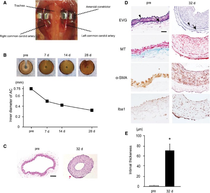
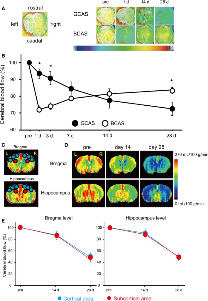
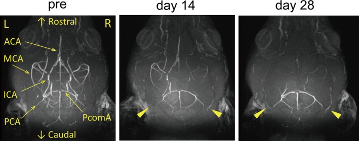
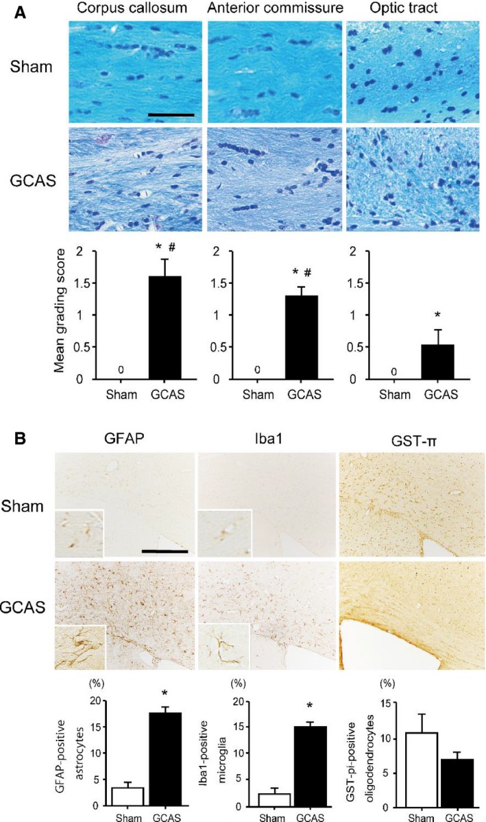
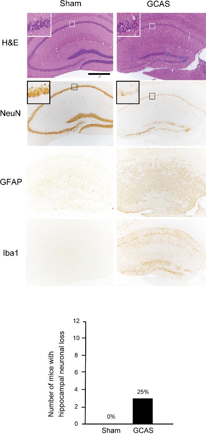
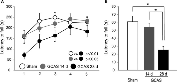
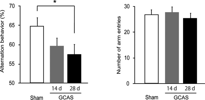
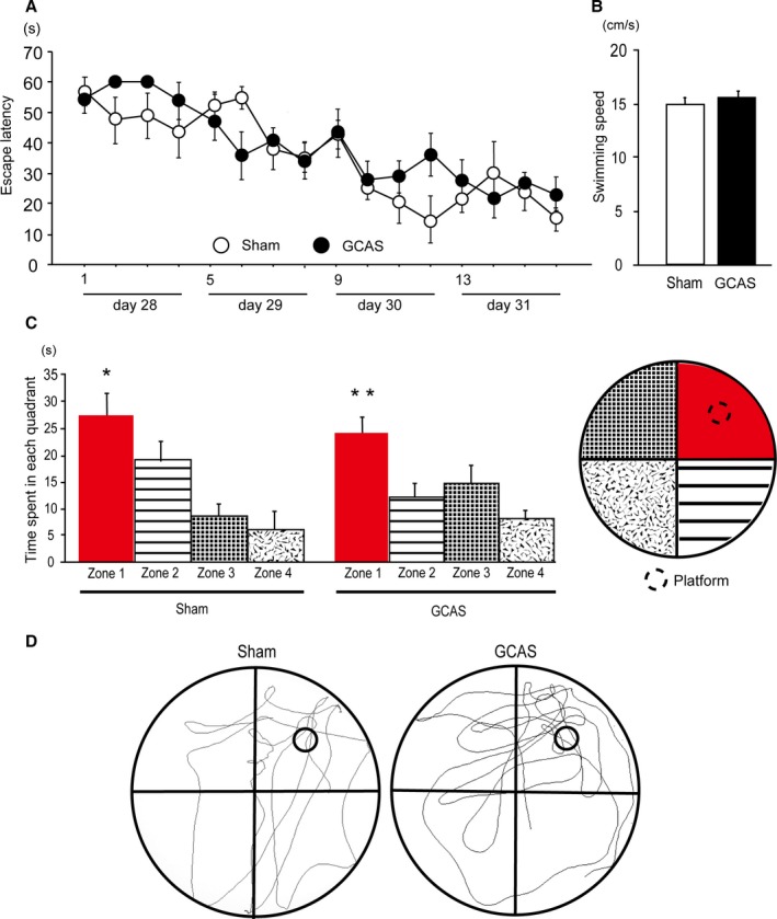
Methods and results: Adult C57BL/6J male mice were subjected to gradual common carotid artery stenosis (GCAS) surgery by using an ameroid constrictor vessel-constricting device with an inner diameter of 0.75 mm. The common carotid arteries narrowed gradually after gradual constriction of ameroid constrictors over 28 days after GCAS, with subsequent 79.3% area stenosis as a result of smooth muscle cell proliferation and macrophage infiltration in the tunica intima. The 28-day survival rate was 91%. Arterial spin labeling demonstrated gradual and continuous reduction of cortical and subcortical CBF (ratio to the preoperative value) to 54.6% and 51.5%, respectively, over 28 days. However, magnetic resonance angiography showed increment of collateral flow signals in the leptomeningeal artery. Rarefaction and proliferation of astrocytes and microglia, with loss of oligodendrocytes, were found in the white matter at 32 days. Hippocampal neuronal loss was observed in only 25% of GCAS mice, consistent with lack of abnormalities in the Morris water maze test. The rotarod test showed motor impairment, and the Y-maze test showed working memory deficits.

Conclusions: The GCAS model successfully generated gradual and continuous CBF reduction over 28 days, with replication of key histological, radiological, and behavioral features associated with cerebral hypoperfusion leading to VCI.

Keywords: ameroid constrictor; carotid artery stenosis; mouse; subcortical ischemic vascular dementia; vascular cognitive impairment.


Figures

Figure 1
Figure 1
Ameroid constrictors (ACs) induce intimal thickening and luminal stenosis in bilateral common carotid arteries (CCAs). A, An image showing surgical implantation of the ACs on the bilateral CCAs. B, Representative images of ACs at indicated time points showing that a central hole (lumen) gradually narrows. Lower graph shows temporal change of inner diameter of ACs (n=2). C, Representative images of the preoperative (left) and postoperative (right) transverse sections of CCA stained with hematoxylin–eosin. D, Representative images of the preoperative (left) and postoperative (right) transverse sections of CCA stained with elastica van Gieson (EVG), Masson trichrome (MT), α–smooth muscle actin (α‐SMA), and ionized calcium binding adaptor molecule 1 (Iba1) stains. Gradual CCA stenosis (GCAS) surgery induced substantial intimal thickening at 32 days after surgery, although the tunica intima of preoperative CCAs was thin. Arrowheads indicate internal elastic lamina, and arrows external elastic lamina. Scale bars indicate 50 μm (C) and 20 μm (D). E, Histogram showing intimal thickness of CCAs before and after GCAS (n=3). *P<0.01.
Figure 2
Figure 2
Temporal profiles of cerebral blood flow (CBF) in mice subjected to gradual common carotid artery stenosis (GCAS). A and B, Representative images of laser speckle flowmetry (A) and temporal profiles of cortical surface CBF (B) in mice subjected to GCAS (n=12) and bilateral CCA stenosis with microcoils (bilateral CCA stenosis [BCAS]; n=7). The levels of cortical surface CBF estimates at indicated time points (before, and 1, 3, 7, 14, and 28 days after each surgery) are shown as percentage of the baseline CBF. Two groups were not significantly different in 2‐way repeated‐measures ANOVA. *P<0.01, GCAS vs BCAS at indicated each time point. C, Regions of interest (ROIs) used for measurement of CBF images obtained from arterial spin labeling (ASL) magnetic resonance perfusion imaging. The CBF values in cerebral cortical area were calculated from the 6 circular ROIs in blue and those in the subcortical area from the 2 circular ROIs in red. D, Representative multislice coronal CBF images obtained from ASL at the bregma and hippocampal levels pre‐GCAS surgery and at 14 and 28 days after GCAS surgery. E, Their temporal profiles of CBF in the cortical (blue) and subcortical areas (red) of GCAS mice (n=4). Two groups were not significantly different at the bregma and hippocampus levels.
Figure 3
Figure 3
Gradual reductions of intracranial arterial flow signals after gradual common carotid artery stenosis (GCAS). Representative images of intracranial arterial flow signals obtained from 7‐T brain magnetic resonance angiography before GCAS (left), and at 14 days (middle) and 28 days (right) after GCAS. Arrowheads indicate gradual increment of collateral flow signals from posterior cerebral artery (PCA) to anterior circulation. ACA indicates anterior cerebral artery; ICA, internal carotid artery; MCA, middle cerebral artery; PcomA, posterior communicating artery.
Figure 4
Figure 4
Hypoperfusive brain injury evident in the white matter (WM). A, Representative photomicrographs of the Klüver–Barrera staining of the paramedial part of the corpus callosum (CC), anterior commissure, and optic tract from the brain of sham‐surgery and gradual common carotid artery stenosis (GCAS) mice at 32 days after each surgery. Lower line graphs show temporal profiles of the grade of WM changes in the respective WM regions. B, Representative photomicrographs of immunostain for glial fibrillary acidic protein (GFAP), ionized calcium binding adaptor molecule 1 (Iba1), and glutathione‐S‐transferase‐π (GST‐π) at the paramedial part of CC from the brain of the sham‐surgery and GCAS mice at 32 days after each surgery. Lower line graphs show temporal profiles of the %numerical densities of the GFAP‐positive astrocytes, Iba1‐positive microglia, and GST‐π–positive oligodendrocytes from the brains of sham‐surgery and GCAS mice at day 32 after each surgery. Scale bars indicate 50 μm in (A) and 200 μm in (B). *P<0.01 vs sham; # P<0.01 vs optic tract.
Figure 5
Figure 5
Only 25% of mice subjected to gradual common carotid artery stenosis (GCAS) surgery showed hippocampal neuronal loss. A GCAS mouse showed hippocampal neuronal loss in hematoxylin–eosin (H&E) stain and NeuN immunohistochemistry. Hippocampal neuronal loss was surrounded by GFAP‐positive astrocytes and Iba1‐positive microglia. The lower line histogram shows number of mice with hippocampal neuronal loss. Hippocampal neuronal loss was seen in only 3 of 12 GCAS mice and no sham‐surgery mice (sham‐surgery, n=10; GCAS mice, n=12).
Figure 6
Figure 6
Impaired motor performance of gradual common carotid artery stenosis (GCAS) mice in rotarod and wire hang tests. A, Motor coordination and balance tested with 5 consecutive trials of rotarod test on days 14 and 28. B, Neuromuscular strength tested with wire hang test on days 14 and 28. *P<0.01. All the behavioral studies were performed in the GCAS mice at day 14 (n=8) and day 28 (n=12), and sham‐surgery mice (n=10).
Figure 7
Figure 7
Impaired working memory of gradual common carotid artery stenosis (GCAS) mice in Y‐maze test. Spatial working memory and spontaneous activity were tested with Y‐maze test on days 14 and 28. GCAS mice at day 28 showed spatial working memory impairment but no spontaneous activity alterations. All the behavioral studies were performed in the GCAS mice at day 14 (n=8) and day 28 (n=12), and sham‐surgery mice (n=10). *P<0.05.
Figure 8
Figure 8
Preserved reference learning and memory of gradual common carotid artery stenosis (GCAS) mice in the Morris water maze test. A, Time course of escape latency from day 1 to 4 of sham‐surgery and GCAS mice in the acquisition phase is shown; no significant difference was found between the 2 groups. B, Mean swimming speed in the acquisition phase was not different between the 2 groups. C, Histogram showing the time spent in each quadrant in the probe trial. *P<0.05 vs Zones 2, 3, and 4. **P<0.01 vs Zones 2, 3, and 4. D, Representative images of traces of the swimming paths of 2 groups in the probe trial. The Morris water maze test was performed in the GCAS mice (n=12) and the sham‐surgery mice (n=10).

References

    1. Kapourchali FR, Surendiran G, Chen L, Uitz E, Bahadori B, Moghadasian MH. Animal models of atherosclerosis. World J Clin Cases. 2014;2:126–132. - PMC - PubMed
    1. Hattori Y, Kitamura A, Nagatsuka K, Ihara M. A novel mouse model of ischemic carotid artery disease. PLoS One. 2014;9:e100257. - PMC - PubMed
    1. Iadecola C. The pathobiology of vascular dementia. Neuron. 2013;80:844–866. - PMC - PubMed
    1. Kalaria RN, Akinyemi R, Ihara M. Does vascular pathology contribute to Alzheimer changes? J Neurol Sci. 2012;322:141–147. - PubMed
    1. Roman GC. Vascular dementia: distinguishing characteristics, treatment, and prevention. J Am Geriatr Soc. 2003;51:S296–S304. - PubMed

Publication types

MeSH terms