. 2016 Apr;22(2):349-60.
doi: 10.1017/S1431927616000088. Epub 2016 Feb 23.

2-Photon Characterization of Optical Proteolytic Beacons for Imaging Changes in Matrix-Metalloprotease Activity in a Mouse Model of Aneurysm

Affiliations

2-Photon Characterization of Optical Proteolytic Beacons for Imaging Changes in Matrix-Metalloprotease Activity in a Mouse Model of Aneurysm

Darren G Haskett et al. Microsc Microanal. 2016 Apr.

Abstract

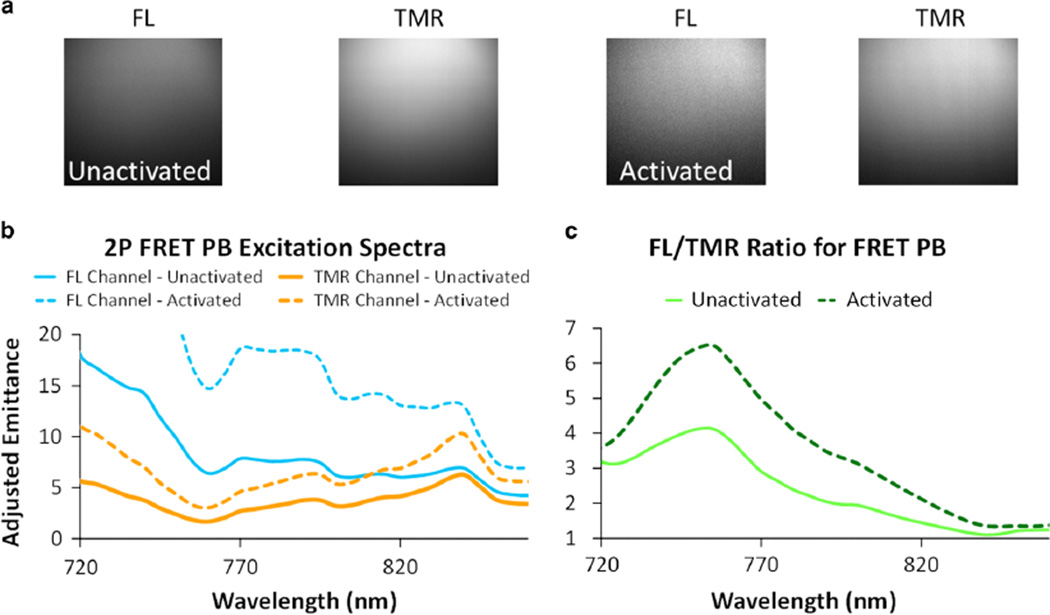
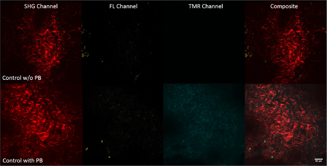
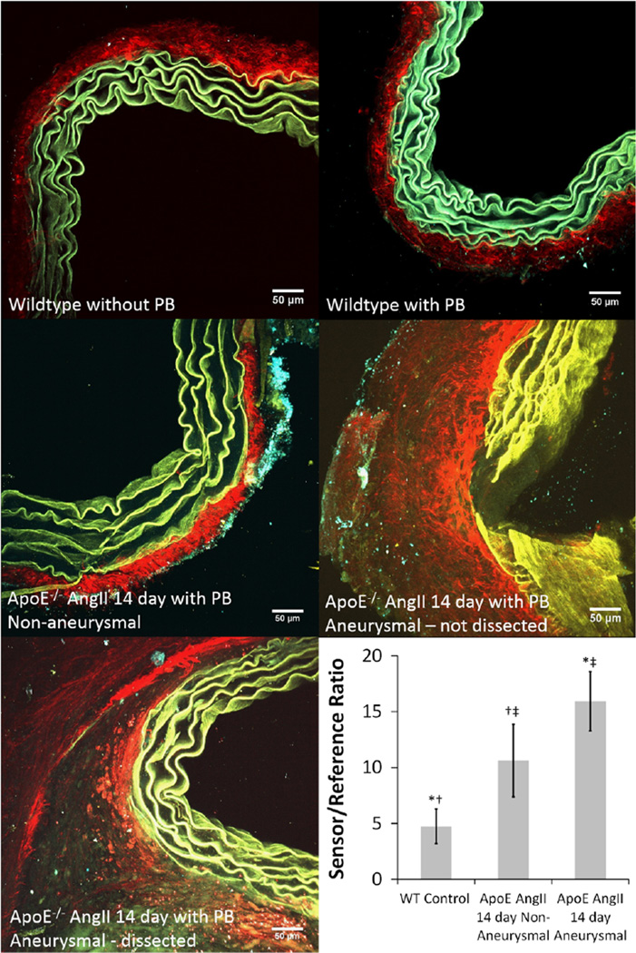
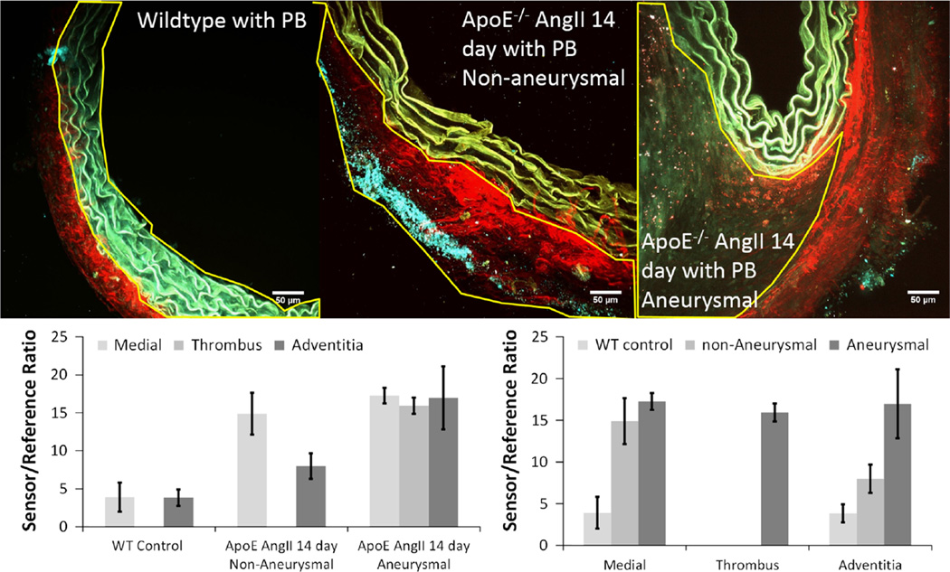
Abdominal aortic aneurysm is a multifactorial disease that is a leading cause of death in developed countries. Matrix-metalloproteases (MMPs) are part of the disease process, however, assessing their role in disease initiation and progression has been difficult and animal models have become essential. Combining Förster resonance energy transfer (FRET) proteolytic beacons activated in the presence of MMPs with 2-photon microscopy allows for a novel method of evaluating MMP activity within the extracellular matrix (ECM). Single and 2-photon spectra for proteolytic beacons were determined in vitro. Ex vivo experiments using the apolipoprotein E knockout angiotensin II-infused mouse model of aneurysm imaged ECM architecture simultaneously with the MMP-activated FRET beacons. 2-photon spectra of the two-color proteolytic beacons showed peaks for the individual fluorophores that enable imaging of MMP activity through proteolytic cleavage. Ex vivo imaging of the beacons within the ECM revealed both microstructure and MMP activity. 2-photon imaging of the beacons in aneurysmal tissue showed an increase in proteolytic cleavage within the ECM (p<0.001), thus indicating an increase in MMP activity. Our data suggest that FRET-based proteolytic beacons show promise in assessing MMP activity within the ECM and will therefore allow future studies to identify the heterogeneous distribution of simultaneous ECM remodeling and protease activity in aneurysmal disease.

Keywords: AAA; ApoE; FRET; MMP; multiphoton.


Figures

Figure 1
Figure 1
Schematic illustrations of reference compounds and proteolytic beacon (PB) showing (left to right) the polyamidoamine (PAMAM) dendrimer backbone with reference tetramethylrhodamine (TMR) fluorophores, PAMAM dendrimer backbone with peptide linker and sensor fluorescein (FL) fluorophores, and the Förster resonance energy transfer (FRET)-based PB with both reference TMR and peptide-attached sensor FL fluorophores.
Figure 2
Figure 2
(Left) Spectra from fluorescein polyamidoamine (FL-PAMAM) dendrimer excited by 485 nm light and emitting a peak around 520 nm, the tetramethylrhodamine (TMR)-PAMAM dendrimer shows greatly reduced fluorescence at its peak emission (590 nm), whereas the proteolytic beacon (PB) shows minimal FL fluorescence but strong fluorescence corresponding with TMR indicating efficient Förster resonance energy transfer (FRET) in the uncleaved PB. (Right) Emission spectra (ex 490 nm) of PB after 2-h incubation with varying concentrations of trypsin (as indicated) showing enhanced FL sensor fluorescence with increasing trypsin indicating cleavage of PBs in vitro.
Figure 3
Figure 3
(Left) Sensor/reference ratios for single-photon measurements of kinetics of the BR2 peptide proteolytic beacons (PBs) at multiple concentrations (U/ml) incubated in ~72 pMoles PB/ml in reaction buffer for up to 3 h. (Right) The sensor/reference ratio at 120 min plotted against matrix-metalloprotease (MMP) concentration representing the reaction progress enzyme kinetics.
Figure 4
Figure 4
a: 2-photon (2P) images of the fluorescence of proteolytic beacons (PBs) in suspension in the fluorescein (FL) and tetramethylrhodamine (TMR) channels for both unactivated and activated PBs. Activation is evidenced by the increase in the mean intensity in the FL channel for the activated PBs and the enhanced FL/TMR ratio. b: 2P spectral excitation sweeps for the PB before (solid lines) and after (dotted lines) protease activation for both the FL (blue) and TMR (orange) emission channels. c: Changes in the FL/TMR (sensor/reference) 2P fluorescence ratio illustrates a peak around 760 nm excitation that increases significantly with protease activation. FRET, Förster resonance energy transfer.
Figure 5
Figure 5
(Top row) Representative 2-photon (2P) single-slice images of the adventitia for second harmonic generation (SHG), fluorescein (FL), tetramethylrhodamine (TMR), and overlaid channels (from left to right) before incubation with proteolytic beacons (PBs) for a wildtype control mouse depicting collagen signal (red), whereas signal in the FL (yellow) and TMR (cyan) channels is virtually absent. (Bottom row) Representative 2P single-slice images of the adventitia for SHG, FL, TMR, and overlaid (from left to right) channels after 16 h of incubation with PBs for a wildtype control mouse depicting collagen signal (red), and signal in the FL (yellow) and TMR (cyan) channels. The dynamic range of the FL channel and TMR channel of the top and bottom rows are 0–64 and 0–255, respectively, in order to illustrate the difference in signal in the two channels within the adventitia before and after the addition of PBs.
Figure 6
Figure 6
(Top row) Representative 2-photon (2P) single-slice images of the adventitia for second harmonic generation (SHG), fluorescein (FL), tetramethylrhodamine (TMR), and overlaid (from left to right) channels after being incubated with proteolytic beacons (PBs) for a wildtype control mouse aorta depicting collagen signal (red), with signal in the FL (yellow) and TMR (cyan) channels indicating MMP activity. (Bottom row) Representative 2P single-slice images of the adventitia for SHG, FL, TMR, and overlaid channels after 16 h of incubation with PBs for an apolipoprotein E knockout 14-day angiotensin II-infused remodeled mouse aorta depicting collagen signal (red), with increased signal in the FL (yellow) and TMR (cyan) channels. Notice that where SHG is present, autofluorescence is absent, so in the corresponding images, the fluorescence is being generated primarily by the PBs.
Figure 7
Figure 7
2-photon maximum intensity projections for cross-sectional image stacks of suprarenal aortas from wildtype (WT) mouse without proteolytic beacons (PBs) (top left), WT mouse after 16-h immersion in PBs (top right), apolipoprotein E knockout (ApoE−/−) 14-day angiotensin II (AngII)-infused nonaneurysmal (middle left), ApoE−/− 14-day AngII-infused non-dissected aneurysmal (middle right), and ApoE−/− 14-day AngII-infused dissected aneurysmal (bottom left) aorta using 4 µm steps up to 100 µm. Images are an overlay of three channels: second harmonic generation (red) primarily depicting collagen, fluorescein (FL) channel (yellow) signifying autofluorescence of the elastin in the medial layer of the aorta and the cleaved FL fluorophore, and tetramethylrhodamine (TMR) channel (cyan) signifying autofluorescence of the elastin in the medial layer of the aorta and the remaining uncleaved PBs still undergoing Förster resonance energy transfer. The mean intensity in the FL and TMR channels for each maximum intensity projection for every slice from each specimen was used to give the sensor/reference ratio (bottom right) and indicates an increase in matrix-metalloprotease (MMP) activity for both ApoE−/− AngII-infused non-aneurysmal and aneurysmal aortas (*, †p < 0.001). It was also found that there was a significant increase in MMP activity between ApoE−/− AngII-infused non-aneurysmal and aneurysmal aortas (‡p = 0.019).
Figure 8
Figure 8
2-Photon maximum intensity projections for cross-sectional image stacks of suprarenal aortas from wildtype (WT) mouse (top left), apolipoprotein E knockout (ApoE−/−) 14-day angiotensin II (AngII)-infused non-aneurysmal (top middle), and ApoE−/− 14-day AngII-infused dissected aneurysmal aortas (top right) with regions of interest (ROIs) outlined indicating spatial distinctions between the media (top left), adventitia (top middle), and thrombus (top right). Images are an overlay of three channels: second harmonic generation (red) primarily depicting collagen, fluorescein (FL) channel (yellow) signifying autofluorescence of the elastin in the medial layer of the aorta and the cleaved FL fluorophore, and tetramethylrhodamine (TMR) channel (cyan) signifying the autofluorescence of the elastin in the medial layer of the aorta and the remaining uncleaved PBs still undergoing Förster resonance energy transfer. Sensor/reference ratios (bottom row) for the ROIs based on region and groups. Ratios for each region are comparable within the WT control and AopE−/− AngII-infused aneurysmal groups; however, there was a significant difference between regions within the AopE−/− AngII-infused non-aneurysmal group (p = 0.001).

References

    1. Bal U, Andresen V, Baggett B, Utzinger U. Intravital confocal and two-photon imaging of dual-color cells and extracellular matrix mimics. Microsc Microanal. 2013;19:201–212. - PMC - PubMed
    1. Bartoli MA, Parodi FE, Chu J, Pagano MB, Mao D, Baxter BT, Buckley C, Ennis TL, Thompson RW. Localized administration of doxycycline suppresses aortic dilatation in an experimental mouse model of abdominal aortic aneurysm. Ann Vasc Surg. 2006;20:228–236. - PubMed
    1. Baxter BT, Pearce WH, Waltke EA, Littooy FN, Hallett JW, Jr, Kent KC, Upchurch GR, Jr, Chaikof EL, Mills JL, Fleckten B, Longo GM, Lee JK, Thompson RW. Prolonged administration of doxycycline in patients with small asymptomatic abdominal aortic aneurysms: Report of a prospective (Phase II) multicenter study. J Vasc Surg. 2002;36:1–12. - PubMed
    1. Baxter BT, Terrin MC, Dalman RL. Medical management of small abdominal aortic aneurysms. Circulation. 2008;117:1883–1889. - PMC - PubMed
    1. Cassis LA, Gupte M, Thayer S, Zhang X, Charnigo R, Howatt DA, Rateri DL, Daugherty A. ANG II infusion promotes abdominal aortic aneurysms independent of increased blood pressure in hypercholesterolemic mice. Am J Physiol Heart Circ Physiol. 2009;296:H1660–H1665. - PMC - PubMed

Publication types

Substances