

doi: 10.1038/srep21377.
T cell-specific inactivation of mouse CD2 by CRISPR/Cas9
Affiliations
- PMID: 26903281
- PMCID: PMC4763270
- DOI: 10.1038/srep21377
Item in Clipboard
T cell-specific inactivation of mouse CD2 by CRISPR/Cas9
Sci Rep.
.
Abstract
The CRISPR/Cas9 system can be used to mutate target sequences by introduction of double-strand breaks followed by imprecise repair. To test its use for conditional gene editing we generated mice transgenic for CD4 promoter-driven Cas9 combined with guide RNA targeting CD2. We found that within CD4(+) and CD8(+) lymphocytes from lymph nodes and spleen 1% and 0.6% were not expressing CD2, respectively. T cells lacking CD2 carryied mutations, which confirmed that Cas9 driven by cell-type specific promoters can edit genes in the mouse and may thus allow targeted studies of gene function in vivo.
Figures
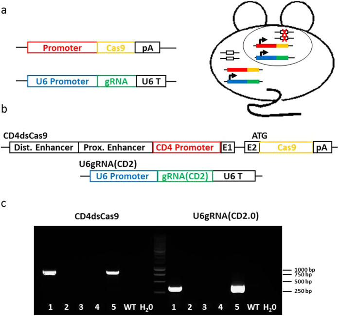
(a) Scheme of the concept of conditional gene editing. In the Cas9 driver strain, the nuclease is placed under control of a cell type or lineage specific promoter. The gRNA construct is driven by the ubiquitous U6 promoter. Both transgenes are co-injected into oocytes. In double-transgenic animals, cell-type specific gene deletions are induced. (b) Scheme of constructs used for the CD4dsCas9/U6gRNA(CD2) mouse strain. The two used linearized plasmids are shown. First, distal and proximal enhancer, CD4 promoter followed by exon 1, part of exon 2 and Cas9 with a PolyA at the end. Second, the U6 promoter driven gRNA specific for CD2 followed by the U6 terminator (U6 T). (c) PCR analysis of tail biopsies for presence of CD4dsCas9 (835 bp amplicon) (lanes 1 and 5) and U6gRNA(CD2.0) (407 bp amplicon) (lanes 1 and 5) by PCR. DNA from a wildtype (WT) mouse as well as H20 were run as a negative control. 2, 3 and 4 were non-transgenic litter mates.
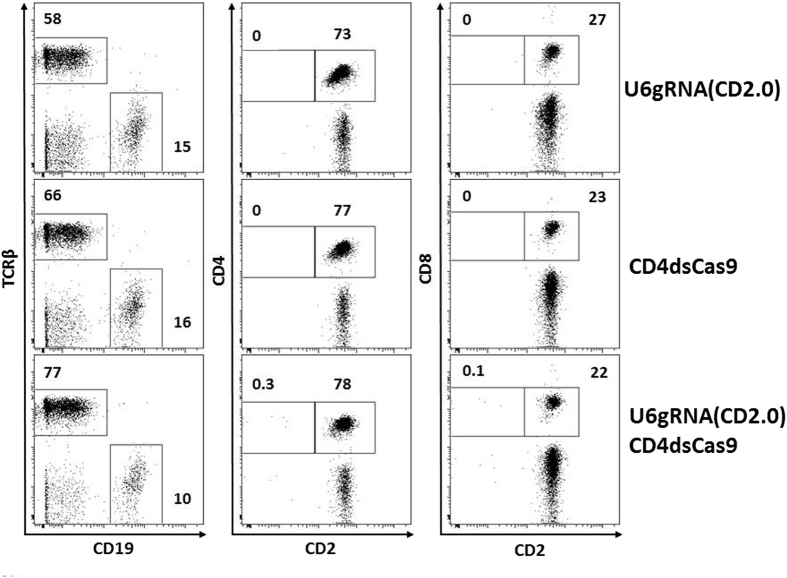
Analysis of blood lymphocytes of the indicated transgenic mice by flow cytometry. Shown are live cells within a lymphocyte gate. CD4 and CD8 cells are additionally gated on TCRβ. The percentages of cells found within the marked gates of the dot plot analysis are shown.
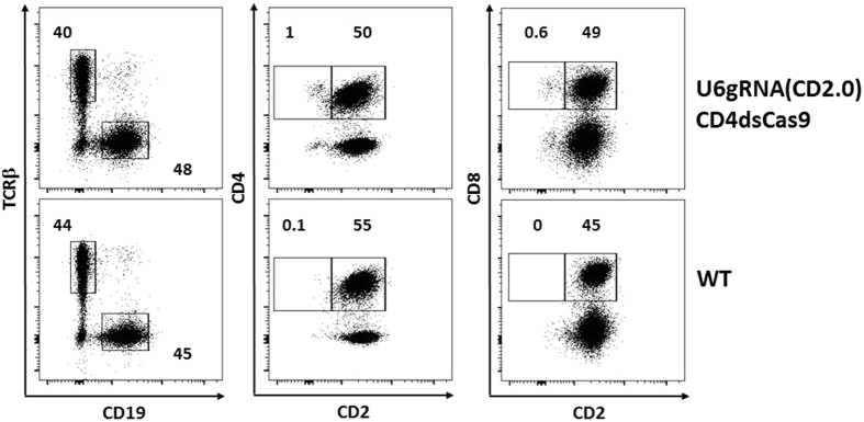
Flow cytometric analysis of pooled lymphocytes from lymph nodes and spleens of two double transgenic and wildtype mice. Shown are live cells within a lymphocyte gate. The four plots on the right are additionally gated on TCRβ+ cells. The percentages of cells found within the marked gates of the dot plot analysis are indicated.
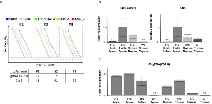
(a) Serial dilutions of genomic DNA from three F1 offspring of founder #5 (Fig. 1c) were analyzed by real-time PCR specific for sequences within the indicated (trans)genes. A regression analysis was performed to calculate the copy number of the transgenes in each individual mouse, as indicated in the table. (b) Relative expression of CD4-Cas9 transgenic and endogenous CD4 mRNA. Transgenic splenic B cells, T cells and thymocytes as well as wildtype thymocytes were magnetically enriched and analyzed by RT-PCR. The CD4-Cas9 product is spanning the intron between CD4 Exon 1 and Cas9 ORF. Cxxc1 and Ywhaz served as housekeeping gene. Data was normalized to “dTG T cells Spleen”. (c) Relative expression of gRNA(CD2.0). Splenocytes and thymocytes of three different dTG offspring as well as of a wildtype control were analyzed by RT-PCR. Cxxc1 and Ywhaz served as housekeeping gene. Data was normalized to “dTG Thymus” of the third offspring.
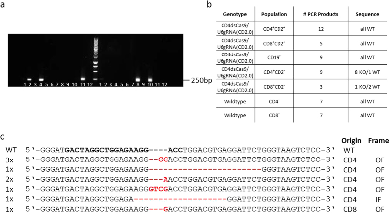
Peripheral blood lymphocytes were surface stained for CD2, CD4, CD8, TCRβ and CD19 and the following populations within live cell and lymphocyte gates single cell-sorted by flow cytometry: TCRβ+ CD4+ CD2+, TCRβ+ CD8+ CD2+, TCRβ+ CD4+ CD2−, TCRβ+ CD8+ CD2−, CD19+. A 253 bp long region including the gRNA target was amplified by two rounds of nested PCR. Products were cloned in pGEM-T and pGEM-Teasy and sequenced. For wildtype controls the single cell PCR products were column-purified and sequenced directly. (a) Agarose gel showing PCR products of single cell amplicons of the target region within the CD2 gene locus from sorted peripheral blood single cells. Lanes 6 and 12 on both sides of the marker are H20 negative controls. (b) Table showing the number of obtained mutations in amplicons of double-transgenic and wildtype cells. (c) Alignment of the obtained sequences from the CD2 gene amplicon. Indicated is the number (left side) and the cell type as well as the outcome of the mutation (OF: out of frame, IF: in frame) (right side) of the respective sequence. – indicates a deletion and red bases insertions.
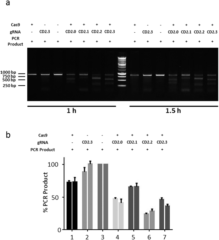
Functional analysis (a) In vitro test of gRNA efficiency. gRNA(CD2.0) as well as three controls (CD2.1,CD2.2, CD2.3) were incubated with a PCR product for the indicated period of time. Digests were separated on an agarose gel. (b) Analysis of the gRNA efficiency. The intensities of the bands resulting from gRNA/Cas9-digested PCR product of three different experiments were analyzed using ImageJ. Shown is the mean and standard error of the mean.
References
-
- Hooper M., Hardy K., Handyside A., Hunter S. & Monk M. HPRT-deficient (Lesch-Nyhan) mouse embryos derived from germline colonization by cultured cells. Nature 326, 292 (1987). - PubMed
-
- Gu H., Marth J. D., Orban P. C., Mossmann H. & Rajewsky K. Deletion of a DNA polymerase beta gene segment in T cells using cell type-specific gene targeting [see comments]. Science 265, 103–106 (1994). - PubMed
-
- Kühn R., Schwenk F., Aguet M. & Rajewsky K. Inducible gene targeting in mice. Science 269, 1427–1429 (1995). - PubMed
MeSH terms
Substances
LinkOut - more resources
Full Text Sources
Other Literature Sources
Molecular Biology Databases
Research Materials
