. 2016 Apr 1;196(7):2933-8.
doi: 10.4049/jimmunol.1501144. Epub 2016 Feb 22.

Cutting Edge: Engineering Active IKKβ in T Cells Drives Tumor Rejection

Affiliations

Cutting Edge: Engineering Active IKKβ in T Cells Drives Tumor Rejection

César Evaristo et al. J Immunol. .

Abstract

Acquired dysfunction of tumor-reactive T cells is one mechanism by which tumors can evade the immune system. Identifying and correcting pathways that contribute to such dysfunction should enable novel anticancer therapy design. During cancer growth, T cells show reduced NF-κB activity, which is required for tumor rejection. Impaired T cell-intrinsic NF-κB may create a vicious cycle conducive to tumor progression and further T cell dysfunction. We hypothesized that forcing T cell-intrinsic NF-κB activation might break this cycle and induce tumor elimination. NF-κB was activated in T cells by inducing the expression of a constitutively active form of the upstream activator IκB kinase β (IKKβ). T cell-restricted constitutively active IKKβ augmented the frequency of functional tumor-specific CD8(+) T cells and improved tumor control. Transfer of constitutively active IKKβ-transduced T cells also boosted endogenous T cell responses that controlled pre-established tumors. Our results demonstrate that driving T cell-intrinsic NF-κB can result in tumor control, thus identifying a pathway with potential clinical applicability.


Figures

Figure 1
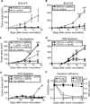
Figure 1. Expression of caIKKβ in T cells improves tumor control in a CD8+ T cell-dependent manner
(A, B) CD4Cre x caIKKβ and littermate control mice were inoculated with 106 B16.SIY (n=3)(A) or B16.F10 (n=3–5)(B) cells s.c. and tumor area was assessed over time. (C-F) CD4Cre x caIKKβ mice were injected with 250µg of control IgG, anti-CD8+anti-CD4 (C, n=3), anti-CD8 (D, n=4) or anti-CD4 (E, n=3) depleting antibodies i.v. at days -7, 0, 7, 14, 21 and 28 relative to inoculation with 106 B16.SIY cells s.c., and tumor area was assessed over time. Tumor growth in littermate controls was assessed for comparison (C, n=5; E, n=1). Depletion efficacy was measured in the blood by flow cytometry (F); circles and squares are plotted against the left and right Y axes, respectively. Data are from 1 (C) or representative of 7 (A) and 2 (B, D-F) independent experiments. Results show mean ± SEM. Differences were determined by two-way ANOVA with Bonferroni’s post-test for multiple comparisons, with *p<0.05, **p<0.01, ***p<0.001.
Figure 2
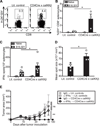
Figure 2. Expression of caIKKβ in T cells results in increased frequency of IFN-γ-producing tumor-specific CD8+ T cells
CD4Cre x caIKKβ or littermate control mice were inoculated with 106 B16.SIY cells. (A, B) Seven days later, the frequency of H2-Kb:SIY-specific CD8+ T cells in the spleen was assessed by flow cytometry. (A) Representative plots. (B) Summarized results (n=3). (C) Number (n=6) and (D) area (n=3) of IFN-γ ELISpots assessed after culture of splenocytes with irradiated tumor cells for 24h. (E) CD4Cre x caIKKβ or littermate control mice were inoculated with 106 B16.SIY cells and injected i.p. with neutralizing anti-IFN-γ or control IgG at days 0, 4, 8, 12, 16 and 20 (n=3–5). Tumor area was analyzed over time. Results show individual mice (B-D) and/or mean ± SEM (B-E) of 1 of 2 independent experiments (B, D, E) or of 2 pooled experiments (C). Differences were determined by Mann-Whitney test (B, C, D) or two-way ANOVA with Bonferroni’s post-test for multiple comparisons (E), with *p<0.05, **p<0.01.
Figure 3
Figure 3. Induction of caIKKβ expression in peripheral T cells is sufficient to enhance control of tumor growth
(A,B) LckCreER x caIKKβ and littermate control mice (Cre-negative-STOPfl/fl-caIKKβ received tamoxifen by gavage (d-7, d-6 and d-4 before inoculation of 106 B16.SIY cells s.c.). (A) Frequency of GFP+ among blood CD4+ or CD8+ T cells at the indicated times relative to the beginning of tamoxifen treatment (n=4–6). (B) Tumor area was analyzed over time (n=9–15). (C) The percentage of transduced T cells was determined in the blood prior to tumor inoculation and at sacrifice (d24) (n=10–13). Results show mean ± SEM and were pooled from 2 (A), 3 (B) and 4 (C) independent experiments. Differences were determined by two-way ANOVA with Bonferroni’s post-test for multiple comparisons (B) or Mann-Whitney test (C), with *p<0.05, **p<0.01, ***p<0.001.
Figure 4
Figure 4. Adoptive transfer of RV-caIKKβ-transduced T cells results in increased control of pre-established tumors
Splenocytes from WT (A-D), 2C/RAG-KO and OTI/RAG-KO (E,F) or Thy1.1 congenic (G,H) mice were transduced with RV-caIKKβGFP or RV–GFP. (A) Single live cells were gated and percent transduced T cells based on GFP expression was assessed by flow cytometry prior to transfer. (B-D) Cells from A, adjusted to contain 0.5–1x10⁶ transduced (GFP+) T cells were adoptively transferred (↓) into WT mice inoculated with 106 B16.SIY cells 7 days earlier. Seven days after transfer, the frequency of H2-Kb:SIY-specific CD8+ T cells in the spleen and tumor was assessed by flow cytometry (n=5) (B) and the frequency of splenocytes that produced IFN-γ when stimulated with SIY peptide was analyzed by ELISpot (n=4) (C). (D) Tumor area was analyzed over time (n=5). (E,F) Cells adjusted to contain 2 × 10⁵ transduced TCR transgenic T cells were transferred into congenic Thy1.2+ WT mice inoculated 7 days earlier with B16.SIY. (E) Tumor growth was analyzed over time (n=5/group). (F) A similar cohort of mice was sacrificed 7 days post-cell transfer (n=5/group) and the percentage of tumor-infiltrating host CD8+ T cells producing IFNγ upon SIY restimulation was determined by flow cytometry. (G,H) Cells adjusted to contain 106 transduced Thy1.1 polyclonal T cells were transferred into congenic Thy1.2+ WT mice that had been inoculated with B16.SIY cells 7 days earlier. Hosts were injected with isotype control (n=5) or anti-Thy1.1 mAb (n=5) on days 14, 16 and 18 post-tumor inoculation. (G) Depletion of Thy1.1+ cells is shown in the spleen and tumor at animal sacrifice. (H) Tumor growth was determined over time. Results show individual mice (B, C, F) and/or mean ± SEM (D, E, G, F)) and are representative of 2 independent experiments. Differences were determined by Mann-Whitney test (B, C, F, G), and by two-way ANOVA with Bonferroni’s post-test for multiple comparisons (D), with *p<0.05, **p<0.01, ***p<0.001. ACT: adoptive cell therapy.

References

    1. Hung K, Hayashi R, Lafond-Walker A, Lowenstein C, Pardoll D, Levitsky H. The central role of CD4(+) T cells in the antitumor immune response. J Exp Med. 1998;188:2357–2368. - PMC - PubMed
    1. Mlecnik B, Tosolini M, Kirilovsky A, Berger A, Bindea G, Meatchi T, Bruneval P, Trajanoski Z, Fridman WH, Pages F, Galon J. Histopathologic-based prognostic factors of colorectal cancers are associated with the state of the local immune reaction. J Clin Oncol. 2011;29:610–618. - PubMed
    1. Harlin H, Meng Y, Peterson AC, Zha Y, Tretiakova M, Slingluff C, McKee M, Gajewski TF. Chemokine expression in melanoma metastases associated with CD8+ T-cell recruitment. Cancer Res. 2009;69:3077–3085. - PMC - PubMed
    1. Ulloa-Montoya F, Louahed J, Dizier B, Gruselle O, Spiessens B, Lehmann FF, Suciu S, Kruit WH, Eggermont AM, Vansteenkiste J, Brichard VG. Predictive gene signature in MAGE-A3 antigen-specific cancer immunotherapy. J Clin Oncol. 2013;31:2388–2395. - PubMed
    1. Zhang L, Conejo-Garcia JR, Katsaros D, Gimotty PA, Massobrio M, Regnani G, Makrigiannakis A, Gray H, Schlienger K, Liebman MN, Rubin SC, Coukos G. Intratumoral T cells, recurrence, and survival in epithelial ovarian cancer. N Engl J Med. 2003;348:203–213. - PubMed

Publication types

MeSH terms