. 2016 Apr 1;143(7):1192-204.
doi: 10.1242/dev.129825. Epub 2016 Feb 22.

Grainyhead-like 2 downstream targets act to suppress epithelial-to-mesenchymal transition during neural tube closure

Affiliations

Grainyhead-like 2 downstream targets act to suppress epithelial-to-mesenchymal transition during neural tube closure

Heather J Ray et al. Development. .

Abstract

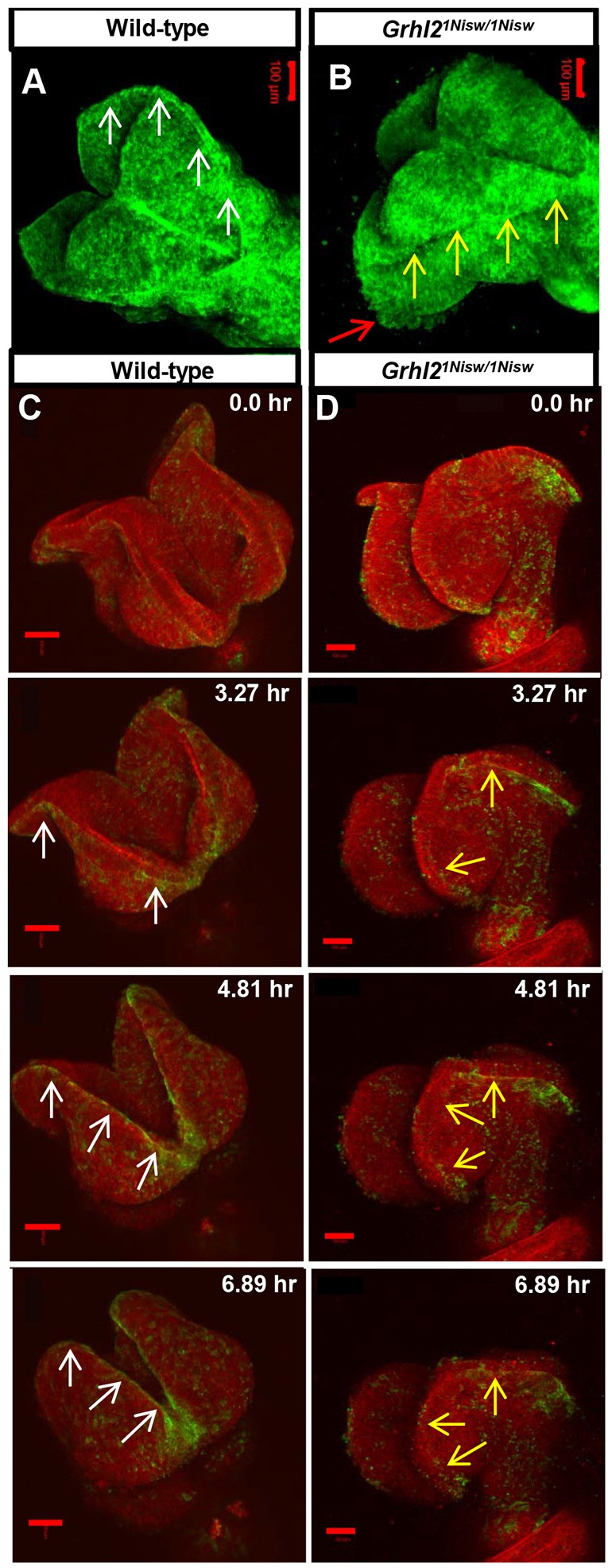
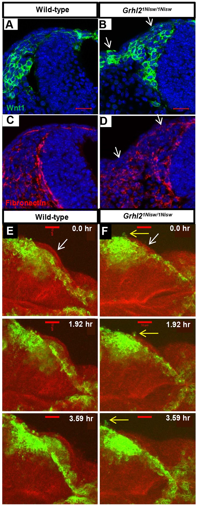
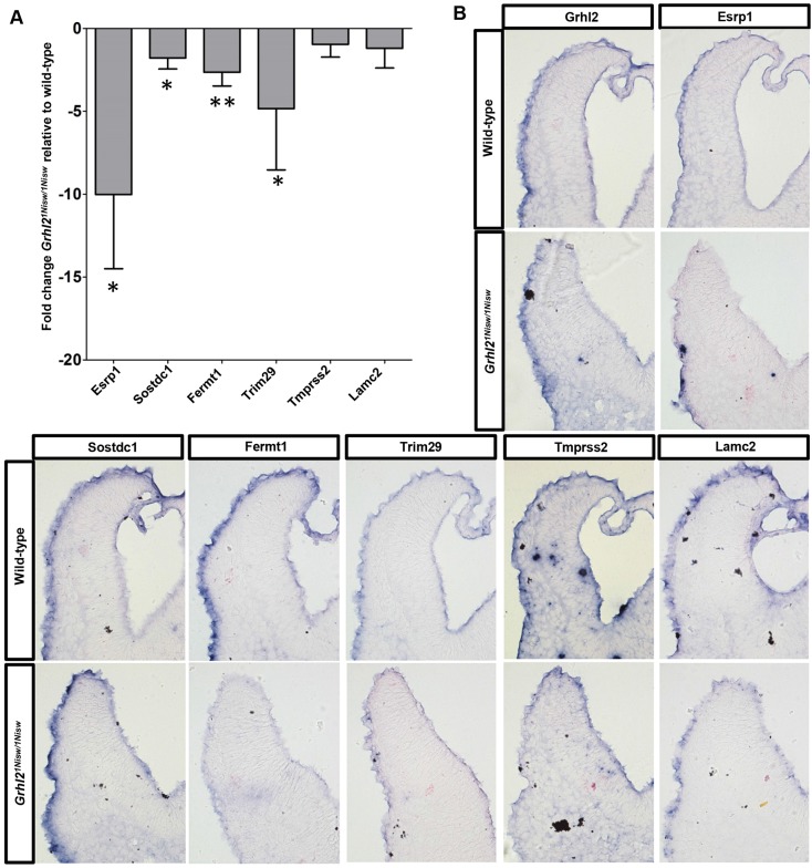
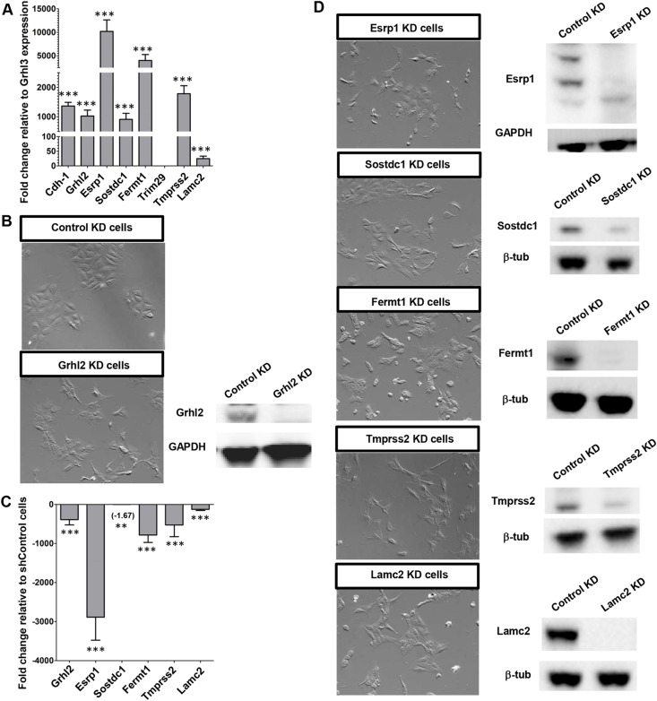
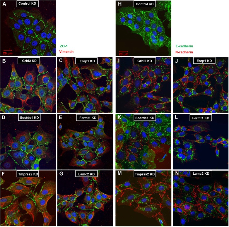
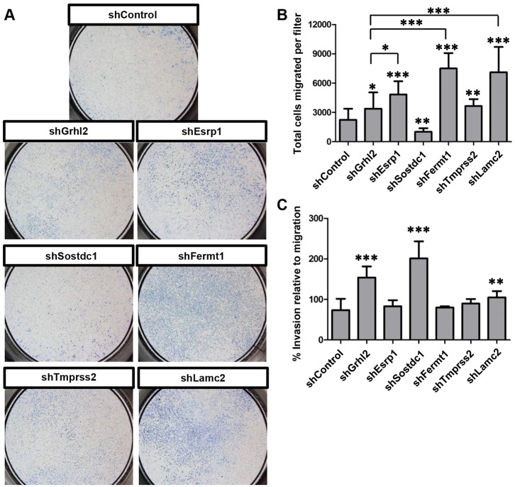
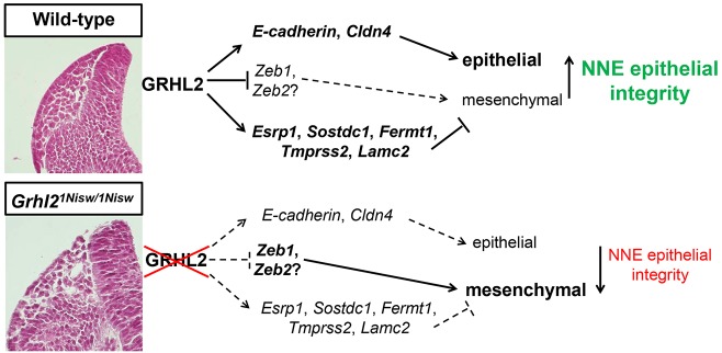
The transcription factor grainyhead-like 2 (GRHL2) is expressed in non-neural ectoderm (NNE) and Grhl2 loss results in fully penetrant cranial neural tube defects (NTDs) in mice. GRHL2 activates expression of several epithelial genes; however, additional molecular targets and functional processes regulated by GRHL2 in the NNE remain to be determined, as well as the underlying cause of the NTDs in Grhl2 mutants. Here, we find that Grhl2 loss results in abnormal mesenchymal phenotypes in the NNE, including aberrant vimentin expression and increased cellular dynamics that affects the NNE and neural crest cells. The resulting loss of NNE integrity contributes to an inability of the cranial neural folds to move toward the midline and results in NTD. Further, we identified Esrp1, Sostdc1, Fermt1, Tmprss2 and Lamc2 as novel NNE-expressed genes that are downregulated in Grhl2 mutants. Our in vitro assays show that they act as suppressors of the epithelial-to-mesenchymal transition (EMT). Thus, GRHL2 promotes the epithelial nature of the NNE during the dynamic events of neural tube formation by both activating key epithelial genes and actively suppressing EMT through novel downstream EMT suppressors.

Keywords: Epithelial-to-mesenchymal transition; Grhl2; Mouse; Neural crest cells; Neural tube closure; Non-neural ectoderm; Trim29.


Conflict of interest statement

Competing interests

The authors declare no competing or financial interests.

Figures

Fig. 1.
Fig. 1.
Grhl2 mutant NNE exhibits loss of epithelial integrity and increased mesenchymal properties. (A-D) H&E staining of transverse sections of cranial neural folds of 13-somite mouse embryos shows tightly associated wild-type NNE (A,C, arrows) versus the loosely associated epithelium and altered cellular phenotype of Grhl2 mutant NNE (B,D, arrows). (E,F) Immunostaining shows regular, punctate ZO-1 (green arrows) and low-level vimentin expression in wild-type NNE (E) versus an irregular ZO-1 expression pattern and aberrant vimentin (red arrows) in Grhl2 mutant NNE (F). (G-J) Quantitation of A-F. (G) NNE breaks in Grhl2 mutants (seven embryos, 50 sections each) are increased compared with wild type (five embryos, 50 sections each). (H,I) The number of ZO-1 puncta per 100 µm shows greater spread and overall smaller mean (H) and the distance between puncta is greater in Grhl2 mutant embryos than in wild type (I). (J) Vimentin expression is increased in Grhl2 mutant NNE compared with wild type. Experiments were performed on four embryos per genotype, ten sections each. Mean±s.d. **P<0.001, ***P<0.0001, Student's t-test. FB, forebrain; HB, hindbrain; RSC, rostral spinal cord; NNE, non-neural ectoderm; WT, wild type. Scale bars: 20 µm.
Fig. 2.
Fig. 2.
Live imaging reveals dynamic alterations to NNE epithelial integrity in Grhl2 mutants. (A,B) Still images from movies of myr-Venus wild-type and Grhl2 mutant embryos (11 somites; dorsal view). Wild-type embryos exhibit a tight line of membrane association at the neural fold tips (A, arrows) and a smooth external epithelial surface, whereas Grhl2 mutant embryos lack the defined membrane association (B, yellow arrows) and have a rough epithelial surface (red arrow) as the neural folds fall away. (C,D) Time series still images from live imaging of closure point 3 of mTmG:Grhl3Cre/+ wild-type and Grhl2 mutant embryos (11 somites). Wild-type NNE cells exhibit tight membrane association at the NE border (C, arrows) as the folds move toward the midline. Neural folds of Grhl2 mutants fall away and do not meet at the midline, and the membrane association at the NNE/NE border is discontinuous and ill-defined (D, arrows). Scale bars: 100 µm.
Fig. 3.
Fig. 3.
Aberrant localization of migrating NCCs reflects loss of NNE epithelial integrity in Grhl2 mutants. (A-D) Fibronectin immunostaining and NCC visualization (mTmG:Wnt1Cre) shows NCC migration between the NE and NNE and an intact basement membrane under the NNE in wild type (A,C), whereas in Grhl2 mutants the NNE layer contains a few NCCs and this correlates with regions of basement membrane disruption (arrows, B,D). Three embryos per genotype were analyzed. (E,F) Time series from live imaging of mTmG:Wnt1Cre/+ wild-type and Grhl2 mutant embryos (11 somites at start; dorsal view) showing NCCs at the edge of the closing neural folds and below the single layer of NNE (E,F, white arrows). In wild-type embryos, NCCs always move internally (E). In Grhl2 mutants, a few NCCs are seen within the NNE layer and they extend cellular processes externally (F, yellow arrows). Scale bars: 20 µm in A,B; 50 µm in E,F.
Fig. 4.
Fig. 4.
Esrp1, Sostdc1, Fermt1, Trim29, Tmprss2 and Lamc2 expression is reduced in Grhl2 mutant embryos. (A) qRT-PCR analysis shows decreased expression of NNE genes in Grhl2 mutant versus wild-type embryos. Experiments were performed in biological quadruplicate with technical quintuplicates. Mean±s.d. *P<0.05, **P<0.01, Student's t-test. (B) In situ hybridization of transverse sections of cranial neural folds shows altered expression of Esrp1, Fermt1, Trim29, Tmprss2 and Lamc2 in Grhl2 mutant NNE. Four embryos per genotype were analyzed in separate technical replicates.
Fig. 5.
Fig. 5.
Loss of Grhl2, Esrp1, Sostdc1, Fermt1, Tmprss2 or Lamc2 in vitro results in a mesenchymal phenotype. (A) qRT-PCR analysis of RNA from IMCD-3 cells shows strong expression of Grhl2 and its targets relative to Grhl3. (B,C) Control KD IMCD-3 (shControl) cells have an epithelial cobblestone morphology, whereas Grhl2 KD (shGrhl2) cells appear mesenchymal and show a significant decrease in expression levels of Grhl2 and its targets as assessed by qRT-PCR. (A,C) Experiments were performed in biological quadruplicate with technical quintuplicates. Mean±s.d. **P<0.01, ***P<0.001, Student's t-test. (D) Cells subject to individual gene KD for Esrp1, Sostdc1, Fermt1, Tmprss2 or Lamc2 exhibit a mesenchymal morphology.
Fig. 6.
Fig. 6.
Loss of Grhl2, Esrp1, Sostdc1, Fermt1, Tmprss2 or Lamc2 expression in IMCD-3 cells causes altered protein expression indicative of an EMT. Immunofluorescence for ZO-1 and vimentin (A-G) or E-cadherin and N-cadherin (H-N) on stable KD cell lines. (A) shControl cells exhibit a highly regular pattern of ZO-1 at the tight junctions between cells, and do not express vimentin. (B) shGrhl2, (C) shEsrp1, (D) shSostdc1, (E) shFermt1, (F) shTmprss2 and (G) shLamc2 cells all show irregular ZO-1 and aberrant vimentin expression patterns. (H) shControl cells exclusively express E-cadherin at the adherens junctions. (I) shGrhl2, (J) shEsrp1, (K) shSostdc1, (L) shFermt1, (M) shTmprss2 and (N) shLamc2 cells all show a switch from E-cadherin to N-cadherin expression. Experiments were performed in triplicate. Scale bars: 20 µm.
Fig. 7.
Fig. 7.
Grhl2, Esrp1, Sostdc1, Fermt1, Tmprss2 and Lamc2 KD cells undergo a functional EMT. (A) Images of whole Transwell filters seeded with IMCD-3 cells in which the specified genes have been subject to KD by shRNA, showing their differential ability to migrate toward a chemoattractant. (B) Quantification of A. (C) Quantification of the total number of KD cells that invaded through a basement membrane relative to migration through the filter alone. Experiments were performed in biological and technical triplicate. Mean±s.d. *P<0.05, **P<0.01, ***P<0.001, Student's t-test.
Fig. 8.
Fig. 8.
Model for GRHL2 regulation of epithelial integrity in NNE. GRHL2 regulates integrity of the NNE through activation of epithelial junctional proteins, direct suppression of ZEB TFs, and activation of several EMT suppressors to enforce the epithelial phenotype of NNE. GRHL2 loss shifts the NNE properties, resulting in loss of epithelial and gain of mesenchymal characteristics.

References

    1. Ai L., Kim W.-J., Alpay M., Tang M., Pardo C. E., Hatakeyama S., May W. S., Kladde M. P., Heldermon C. D., Siegel E. M. et al. (2014). TRIM29 suppresses TWIST1 and invasive breast cancer behavior. Cancer Res. 74, 4875-4887. 10.1158/0008-5472.CAN-13-3579 - DOI - PMC - PubMed
    1. Aue A., Hinze C., Walentin K., Ruffert J., Yurtdas Y., Werth M., Chen W., Rabien A., Kilic E., Schulzke J.-D. et al. (2015). A Grainyhead-Like 2/Ovo-Like 2 pathway regulates renal epithelial barrier function and lumen expansion. J. Am. Soc. Nephrol. 26, 2704-2715. 10.1681/asn.2014080759 - DOI - PMC - PubMed
    1. Aumailley M. and Smyth N. (1998). The role of laminins in basement membrane function. J. Anat. 193, 1-21. 10.1046/j.1469-7580.1998.19310001.x - DOI - PMC - PubMed
    1. Camerer E., Barker A., Duong D. N., Ganesan R., Kataoka H., Cornelissen I., Darragh M. R., Hussain A., Zheng Y.-W., Srinivasan Y. et al. (2010). Local protease signaling contributes to neural tube closure in the mouse embryo. Dev. Cell 18, 25-38. 10.1016/j.devcel.2009.11.014 - DOI - PMC - PubMed
    1. Cecconi F., Alvarez-Bolado G., Meyer B. I., Roth K. A. and Gruss P. (1998). Apaf1 (CED-4 homolog) regulates programmed cell death in mammalian development. Cell 94, 727-737. 10.1016/S0092-8674(00)81732-8 - DOI - PubMed

Publication types

MeSH terms