. 2016 Apr 15;291(16):8805-15.
doi: 10.1074/jbc.M115.705178. Epub 2016 Feb 22.

Major Histocompatibility Complex (MHC) Class I Processing of the NY-ESO-1 Antigen Is Regulated by Rpn10 and Rpn13 Proteins and Immunoproteasomes following Non-lysine Ubiquitination

Affiliations

Major Histocompatibility Complex (MHC) Class I Processing of the NY-ESO-1 Antigen Is Regulated by Rpn10 and Rpn13 Proteins and Immunoproteasomes following Non-lysine Ubiquitination

Richard Golnik et al. J Biol Chem. .

Abstract

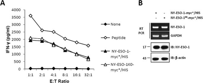
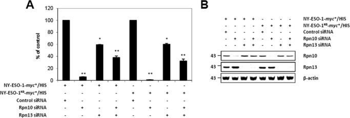
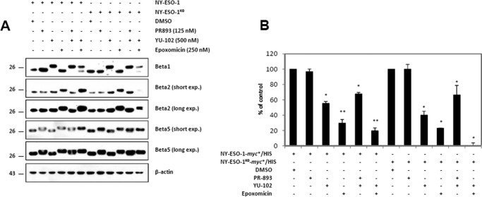
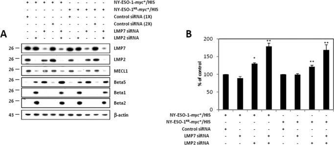
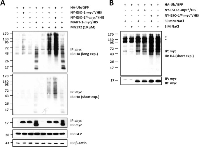
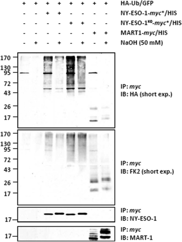
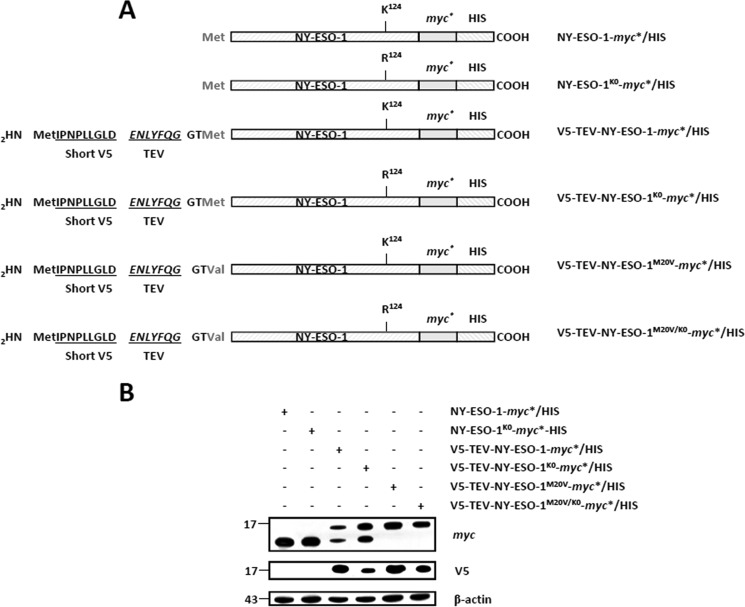
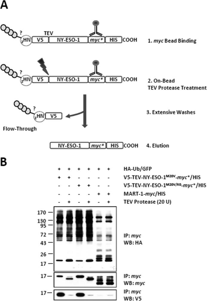
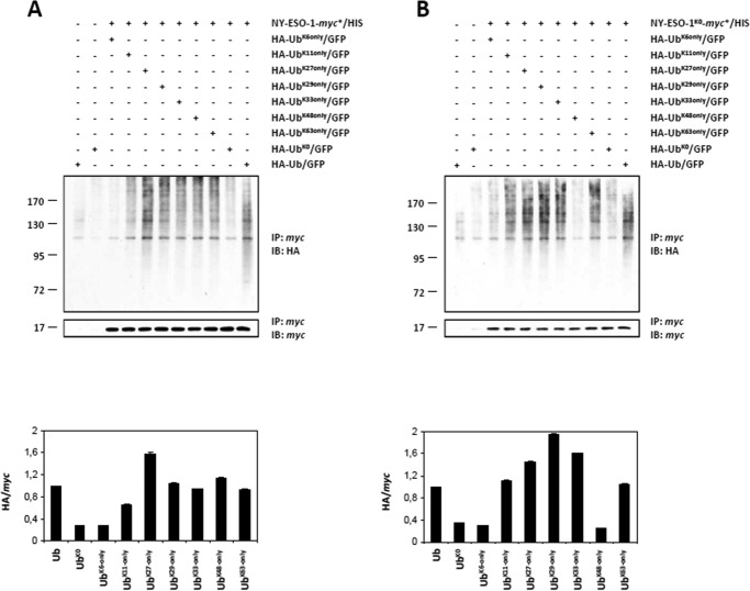
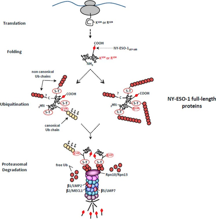
The supply of MHC class I-restricted peptides is primarily ensured by the degradation of intracellular proteins via the ubiquitin-proteasome system. Depending on the target and the enzymes involved, ubiquitination is a process that may dramatically vary in terms of linkages, length, and attachment sites. Here we identified the unique lysine residue at position 124 of the NY-ESO-1 cancer/testis antigen as the acceptor site for the formation of canonical Lys-48-linkages. Interestingly, a lysine-less form of NY-ESO-1 was as efficient as its wild-type counterpart in supplying the HLA-A*0201-restricted NY-ESO-1157-165 antigenic peptide. In fact, we show that the regulation of NY-ESO-1 processing by the ubiquitin receptors Rpn10 and Rpn13 as a well as by the standard and immunoproteasome is governed by non-canonical ubiquitination on non-lysine sites. In summary, our data underscore the significance of atypical ubiquitination in the modulation of MHC class I antigen processing.

Keywords: antigen processing; proteasome; protein degradation; tumor immunology; ubiquitin.


Figures

FIGURE 1.
FIGURE 1.
The unique lysine residue of NY-ESO-1 at position 124 is not essential for NY-ESO-1157–165 presentation. A, HeLa cells were subjected to 24-h transfection with HLA-A*0201 together with either NY-ESO-1 or NY-ESO-1K0. The NY-ESO-1157–165 CTL response was assessed using the CTL clone RG39 specific for NY-ESO-1157–165 at various effector:target ratios as indicated. Internal controls in this experiment consisted of HLA-A*0201-expressing HeLa cells or cells loaded with 10 μm of the 9-mer SLLMWITQV synthetic peptide. After 16 h of co-culture, supernatants were tested for their IFN-γ content by ELISA. The results are expressed as mean ± S.D. of duplicate values. Shown is one representative experiment of three. B, NY-ESO-1 expression was analyzed by RT-PCR (top panel) and Western blotting (bottom panel) as indicated. IB, immunoblot.
FIGURE 2.
FIGURE 2.
The Rpn10 and/or Rpn13 dependences do not vary between NY-ESO-1 and NY-ESO-1K0 for presentation of the NY-ESO-1157–165 antigenic peptide. A, HeLa cells were exposed to control siRNA or siRNA specific against Rpn10 and/or Rpn13 for 3 days prior to subsequent transfection with either NY-ESO-1 or NY-ESO-1K0, as indicated. The NY-ESO-1157–165 CTL response arising from NY-ESO-1 or NY-ESO-1K0 in cells exposed to Rpn10 and/or Rpn13 siRNA was assessed by culturing these cells in the presence of the RG39 CTL clone at a ratio of 1:1. After 16 h of co-culture, supernatants were tested for their IFN-γ content by ELISA. Data are expressed as the percentage relative to cells exposed to control siRNA. Shown is one representative experiment of three. *, p < 0.05; **, p < 0.01 versus control siRNA (Student's t test). B, the knockdown efficiency of Rpn10 and/or Rpn13 in these cells was evaluated by monitoring the steady-state levels of both of these proteins by Western blotting with antibodies specific for Rpn10 and Rpn13.
FIGURE 3.
FIGURE 3.
The generation of the NY-ESO-1157–165 peptide from NY-ESO-1 or NY-ESO-1K0 is mainly driven by the β2 catalytic proteasomal subunit. A, HeLa cells expressing HLA-A*0201 together with either NY-ESO-1 or NY-ESO-1K0 were treated for 2 h with PR-893 (125 nm), YU-102 (500 nm), and epoxomicin (250 nm) individually or in combination, as indicated. Covalent binding of the inhibitors to any of the three standard subunits in cells expressing HLA-A*0201/NY-ESO-1 or HLA-A*0201/NY-ESO-1K0 was monitored by Western blotting using antibodies specific for β1, β2, β5, and β-actin (loading control). DMSO, dimethyl sulfoxide. B, the effects of the PR-893, YU-102, and/or epoxomicin on the NY-ESO-1157–165 CTL responses emerging from NY-ESO-1 or NY-ESO-1K0 were examined in a 16-h CTL assay using RG39 CTL. IFN-γ was measured by ELISA. The results are expressed as percentage relative to cells exposed to dimethyl sulfoxide. Shown is one representative experiment of three. *, p < 0.05; **, p < 0.01 versus dimethyl sulfoxide (Student's t test).
FIGURE 4.
FIGURE 4.
Both NY-ESO-1 and NY-ESO-1K0 are more effective at producing the NY-ESO-1157–165 antigenic peptide in cells lacking the IP-inducible subunits. A, 33/2 cells were exposed to control (once or twice), LMP7, and/or LMP2 siRNA for 2 days before being subjected to a subsequent transfection with NY-ESO-1 or NY-ESO-1K0. Cell extracts were analyzed by Western blotting using antibodies specific for LMP7, LMP2, MECL1, β5, β1, β2, and β-actin (loading control). B, 33/2 cells with a knockdown of LMP7 and/or LMP2 and expressing either NY-ESO-1 or NY-ESO-1K0 were evaluated for their capacity to present the NY-ESO-1157–165 peptide by exposing them to our CTL clone RG39 for 16 h at a effector:target ratio of 8:1. IFN-γ was measured by ELISA. The data are expressed as percentage relative to cells exposed to control siRNA. Shown is one representative experiment of three. *, p < 0.05; **, p < 0.001 versus control siRNA (Student's t test).
FIGURE 5.
FIGURE 5.
The unique lysine residue of NY-ESO-1 at position 124 is not required for NY-ESO-1 ubiquitination. A, whole cell extracts were prepared from HeLa cells co-expressing HA-Ub-GFP together with NY-ESO-1, NY-ESO-1K0, or the MART-1 melanoma antigen (as a control) in the presence or absence of a 6-h treatment with 10 μm MG-132. In vivo ubiquitination of NY-ESO-1 or NY-ESO-1K0 was visualized by pulling down NY-ESO-1 with anti-myc beads, followed by Western blotting using antibodies specific for HA and NY-ESO-1 (which served as a loading control for the pulldown). Cell extracts were subjected to Western blotting analysis using antibodies specific for HA, myc, GFP, and β-actin (loading control) as indicated. IP, immunoprecipitation; IB, immunoblot. B, in vivo ubiquitination of NY-ESO-1 or NY-ESO-1K0 was visualized by precipitating NY-ESO-1 with anti-myc beads prior to five stringent washes with either 50 mm or 3 m NaCl as indicated. After a final wash with 20 mm Tris, the anti-myc-bound material was eluted, and the precipitates were analyzed by Western blotting using antibodies specific for HA and myc. The asterisks represent nonspecific bands. Shown is one representative experiment of two.
FIGURE 6.
FIGURE 6.
Ubiquitination of the NY-ESO-1 and NY-ESO-1K0 tumor antigens occurs on serine and/or threonine residues in HeLa cells. HeLa cells expressing HA-Ub-GFP in combination with NY-ESO-1, NY-ESO-1K0, or the MART-1 melanoma antigen (as a control) were subjected to protein extraction. NY-ESO-1 and/or NY-ESO-1K0 were pulled down by anti-myc beads, and the precipitates were treated with 50 mm NaOH for 25 min at 32 °C or left untreated, as indicated. The negative control for the immunoprecipitation (IP) in this experiment consisted in HeLa cells only transfected with the HA-Ub-GFP construct (first and second lanes). The effect of the alkaline treatment on the ubiquitination of NY-ESO-1, NY-ESO-1K0, and MART-1 was assessed by Western blotting using antibodies specific for HA, ubiquitin, and NY-ESO-1 (loading control). IB, immunoblot.
FIGURE 7.
FIGURE 7.
The expression of an N-terminal V5-tagged version of NY-ESO-1 and/or NY-ESO-1K0 requires the removal of the original NY-ESO-1 start codon. A, sequences of the NY-ESO-1 constructs used in this study. All NY-ESO-1 constructs were C-terminally tagged with a double myc*/HIS epitope. In some experiments, NY-ESO-1-myc*/HIS and NY-ESO-1K0-myc*/HIS were N-terminally tagged with a short 9-mer V5 epitope (IPNPLLGLD) directly upstream of a TEV protease-sensitive site (ENLYFQG). Both V5-TEV-NY-ESO-1-myc*/HIS and V5-TEV-NY-ESO-1K0-myc*/HIS were finally subjected to M20V site-directed mutagenesis to remove the NY-ESO-1 original methionine initiator codon. B, HeLa cells were transfected with each of the six NY-ESO-1 variant for 24 h prior to protein extraction. Whole cell extracts were resolved on 15% SDS-PAGE and subsequently analyzed by Western blotting using anti-myc, anti-V5, and anti-β-actin (loading control) antibodies as indicated. The M20V in both of V5-TEV-NY-ESO-1-myc*/HIS and V5-TEV-NY-ESO-1K0-myc*/HIS prevents the expression of N-terminally untagged NY-ESO-1-myc*/HIS and NY-ESO-1-myc*/HIS, respectively.
FIGURE 8.
FIGURE 8.
The N-terminal −NH2 group of either NY-ESO-1 or NY-ESO-1K0 is not a site of conjugation. A, HeLa cells expressing HA-Ub-GFP in combination with V5-TEV-NY-ESO-1M20V-myc*/HIS or V5-TEV-NY-ESO-1M20V/K0-myc*/HIS were subjected to protein extraction and incubated with μMACS anti-myc beads that were subsequently left untreated or treated with 20 U TEV protease for 150 min at 22 °C as indicated. Extensive washing of the μMACS anti-myc bead-bound material resulted in the elimination of the N terminus and its potential anchored Ub chains in the flow-through fraction. After five washes, the material bound to the μMACS anti-myc beads was eluted. B, the effect of the removal of the V5 N terminus on the ubiquitination of NY-ESO-1 and NY-ESO-1K0 was assessed by Western blotting (WB) using antibodies specific for HA, ubiquitin, V5, and myc (loading control). The negative control for the immunoprecipitation (IP) in this experiment consisted of HeLa cells transfected with the HA-Ub-GFP construct in combination with an expression vector encoding the MART-1 melanoma antigen (fifth and sixth lanes). Shown is one representative experiment of three.
FIGURE 9.
FIGURE 9.
NY-ESO-1 and NY-ESO-1K0 distinguish themselves in terms of poly-Ub chains. A and B, ubiquitination of NY-ESO-1 (A) and NY-ESO-1K0 (B) was examined following overexpression of a HA-tagged wild-type Ub, a Lys-0 HA-tagged Ub (HA-UbK0) devoid of all its seven lysine residues, as well as HA-tagged Lys-6-only, Lys-11-only, Lys-27-only, Lys-29-only, Lys-33-only, Lys-48-only, or Lys-63-only Ub mutants in which all lysine residues were altered to arginine except for lysines 6, 11, 27, 29, 33, 48, and 63, respectively, and as indicated. Ubiquitination of NY-ESO-1 and/or NY-ESO-1K0 was visualized by pulling down NY-ESO-1 with anti-myc beads, followed by Western blotting using antibodies specific for HA and NY-ESO-1 (loading control). Pulldowns with cells overexpressing HA-Ub or HA-UbK0 (first and second lanes) but lacking NY-ESO-1 (A) or NY-ESO-1K0 (B) demonstrate the specificity of the assay. A densitometric analysis of ubiquitinated NY-ESO-1 and NY-ESO-1K0 normalized to pulled-down NY-ESO-1 and NY-ESO-1K0 is plotted (mean ± S.D., n = 2). For both of the ubiquitinated species of NY-ESO-1 and NY-ESO-1K0, the mean of the signals obtained with the wild-type HA-Ub-GFP construct was set at 1. IP, immunoprecipitation; IB, immunoblot.
FIGURE 10.
FIGURE 10.
The processing of NY-ESO-1 relies on atypical ubiquitination occurring on non-lysine sites. The presence of the NY-ESO-1157–165 antigenic peptide at the C terminus of the 180-amino acid-long NY-ESO-1 full-length protein required the protein to be fully synthetized before ubiquitination. Following translation, the full-length NY-ESO-1 protein was subjected to both typical and atypical ubiquitination on Lys-124 and on non-canonical sites (Ser-Thr), respectively. The removal of Lys-124 abrogated the formation of Lys-48-linked poly-Ub chains, which was also accompanied by increased generation of atypical chains. Despite these qualitative and quantities differences, both the wild-type and lysine-less forms of NY-ESO-1 converged on the same regulatory proteasome-mediated MHC class I processing pathway involving the Rpn10 and Rpn13 19S subunits as well as the trypsin-like activity of the β2 standard subunit. This eventually leads to similar production of antigenic peptides, which are equally presented on HLA-A*0201 molecules.

References

    1. Sijts E. J., and Kloetzel P. M. (2011) The role of the proteasome in the generation of MHC class I ligands and immune responses. Cell. Mol. Life Sci. 68, 1491–1502 - PMC - PubMed
    1. Rock K. L., York I. A., Saric T., and Goldberg A. L. (2002) Protein degradation and the generation of MHC class I-presented peptides. Adv. Immunol. 80, 1–70 - PubMed
    1. Murata S., Yashiroda H., and Tanaka K. (2009) Molecular mechanisms of proteasome assembly. Nat. Rev. Mol. Cell Biol. 10, 104–115 - PubMed
    1. Ciechanover A., and Stanhill A. (2014) The complexity of recognition of ubiquitinated substrates by the 26S proteasome. Biochim. Biophys. Acta 1843, 86–96 - PubMed
    1. DeMartino G. N., and Slaughter C. A. (1999) The proteasome, a novel protease regulated by multiple mechanisms. J. Biol. Chem. 274, 22123–22126 - PubMed

Publication types

MeSH terms