. 2016 Apr 8;291(15):8214-30.
doi: 10.1074/jbc.M115.693119. Epub 2016 Feb 22.

Insights into the Effects of Complement Factor H on the Assembly and Decay of the Alternative Pathway C3 Proconvertase and C3 Convertase

Affiliations

Insights into the Effects of Complement Factor H on the Assembly and Decay of the Alternative Pathway C3 Proconvertase and C3 Convertase

Serena Bettoni et al. J Biol Chem. .

Retraction in

Abstract

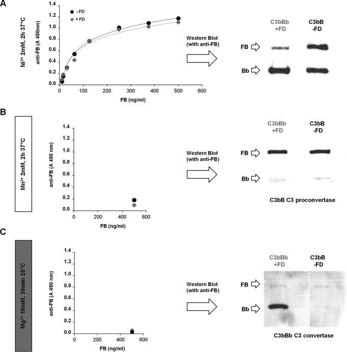
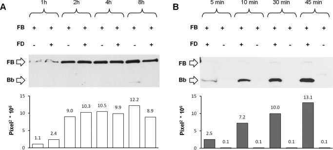
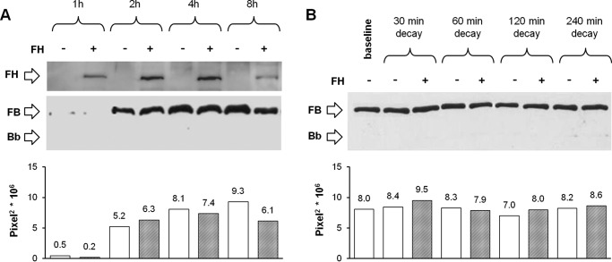
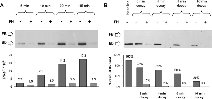
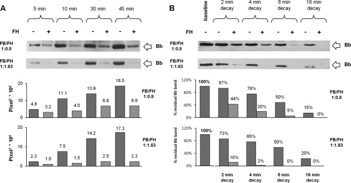
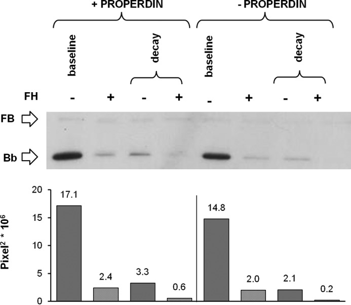
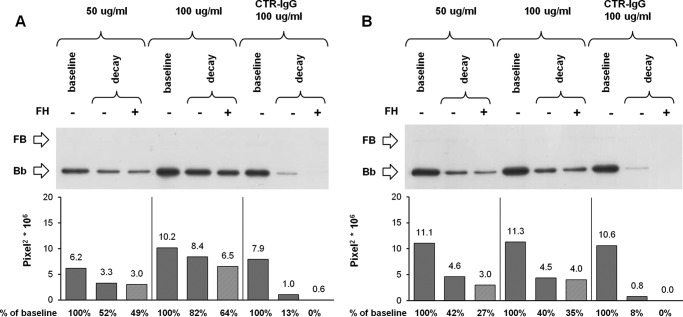
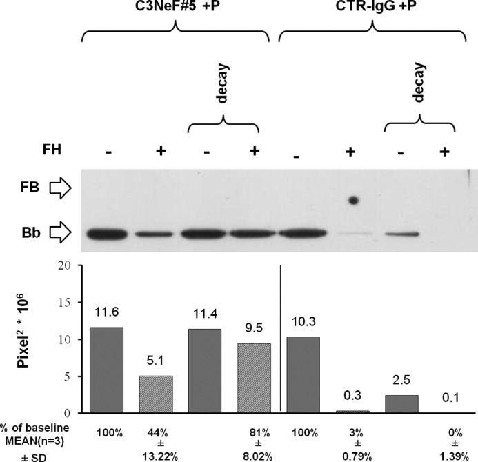
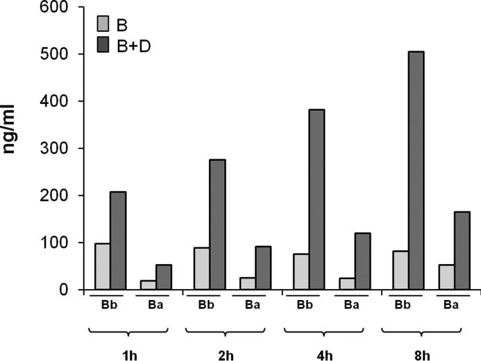
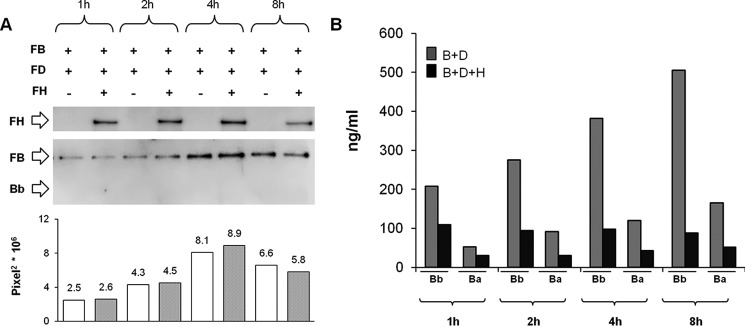
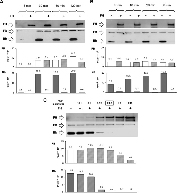
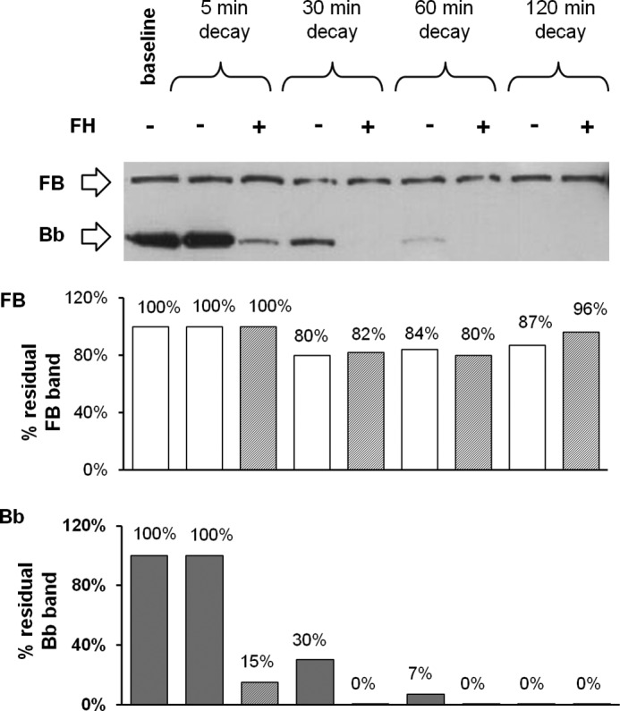
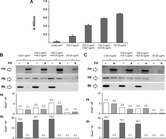
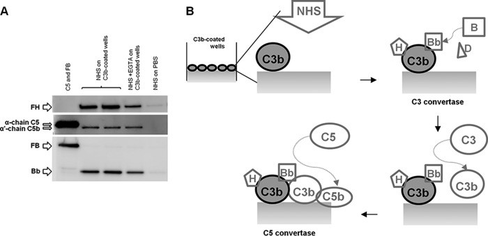
The activated fragment of C3 (C3b) and factor B form the C3 proconvertase (C3bB), which is cleaved by factor D to C3 convertase (C3bBb). Older studies (Conrad, D. H., Carlo, J. R., and Ruddy, S. (1978)J. Exp. Med.147, 1792-1805; Pangburn, M. K., and Müller-Eberhard, H. J. (1978)Proc. Natl. Acad. Sci. U.S.A.75, 2416-2420; Kazatchkine, M. D., Fearon, D. T., and Austen, K. F. (1979)J. Immunol.122, 75-81) indicated that the complement alternative pathway regulator factor H (FH) competes with factor B for C3b binding; however, the capability of FH to prevent C3bB assembly has not been formally investigated. Moreover, in the few published studies FH did not favor C3bB dissociation. Whether FH may affect C3bBb formation from C3bB is unknown. We set up user-friendly assays based on combined microplate/Western blotting techniques that specifically detect either C3bB or C3bBb, with the aim of investigating the effect of FH on C3bB assembly and decay and C3bBb formation and decay. We document that FH does not affect C3bB assembly, indicating that FH does not efficiently compete with factor B for C3b binding. We also found that FH does not dissociate C3bB. FH showed a strong C3bBb decay-accelerating activity, as reported previously, and also exerted an apparent inhibitory effect on C3bBb formation. The latter effect was not fully attributable to a rapid FH-mediated dissociation of C3bBb complexes, because blocking decay with properdin and C3 nephritic factor did not restore C3bBb formation. FH almost completely prevented release of the smaller cleavage subunit of FB (Ba), without modifying the amount of C3bB complexes, suggesting that FH inhibits the conversion of C3bB to C3bBb. Thus, the inhibitory effect of FH on C3bBb formation is likely the sum of inhibition of C3bB conversion to C3bBb and of C3bBb decay acceleration. Further studies are required to confirm these findings in physiological cell-based settings.

Keywords: C3 convertase; C3 nephritic factor; C3 proconvertase; Western blot; alternative pathway complement system; complement; convertase; factor H; magnesium; manganese.


Figures

FIGURE 1.
FIGURE 1.
Experimental design of microplate/WB assays.
FIGURE 2.
FIGURE 2.
Detection of C3bB C3 proconvertase and C3bBb C3 convertase complex formation by ELISA and microplate/WB assays. A, detection of Ni2+-dependent C3bB and C3bBb. C3b-coated microtiter wells were incubated with increasing amounts of FB (0–500 ng/ml) and 2 mm NiCl2 in the absence or in the presence of 25 ng/ml FD and analyzed by ELISA by using an anti-FB antibody (left side). The product of the reaction with 500 ng/ml FB, once detached from the wells, was also analyzed by WB with an anti-FB antibody (right side). B and C, selective formation of C3bB and C3bBb with Mn2+ and Mg2+, respectively. C3b-coated wells were incubated with 500 ng/ml FB in the presence or in the absence of 25 ng/ml FD, 2 mm MnCl2, or 10 mm MgCl2 to obtain C3bB(Mn2+) and C3bBb(Mg2+), respectively. Complexes were detected both by ELISA (left side) and WB (right side). C3bB and C3bBb formation was evaluated by the visualization of the B band (93 kDa) and the Bb band (60 kDa), respectively. One representative experiment of three is shown.
FIGURE 3.
FIGURE 3.
Time course of selective C3bB(Mn2+) C3 proconvertase assembly (A) and C3bBb(Mg2+) C3 convertase formation (B) detected by microplate/WB assays. C3bB(Mn2+) and C3bBb(Mg2+) complexes were obtained by incubating C3b-coated wells at time points indicated with 1000 ng/ml FB in the presence or in the absence of 5 ng/ml FD and 2 mm MnCl2 at 37 °C or 10 mm MgCl2 at 25 °C, respectively. The amount of C3bB assembled and of C3bBb formed was calculated as the intensity of B band (93 kDa) and Bb band (60 kDa), respectively, and results are reported in the bottom graphs as pixel2·106. Results of a representative microplate/WB experiment of n = 3 are shown.
FIGURE 4.
FIGURE 4.
Effect of FH on C3bB(Mn2+) C3 proconvertase assembly (A) and C3bB(Mn2+) C3 proconvertase decay (B) detected by microplate/WB assays. A, time course of C3bB(Mn2+) assembly. The complexes were obtained by incubating C3b-coated wells at 37 °C for 1, 2, 4, and 8 h with 1000 ng/ml FB and 2 mm MnCl2 in the presence or in the absence of 2640 ng/ml FH. FH band (150 kDa) could be visualized in the WB, and a representative image is reported on the top. B, time course of spontaneous and FH-mediated decay of C3bB(Mn2+) assembled in 2 h at 37 °C was evaluated by further incubation with buffer alone or with 2640 ng/ml FH for 30, 60, 120, and 240 min at 37 °C. The amount of C3 proconvertase (B band, 93 kDa) was quantified, and results of corresponding densitometries are reported in the graphs below as pixel2·106. The percentage of residual B band was calculated as the ratio of the densities (in pixel2) of each B band after decay and the corresponding baseline B band density before decay × 100. Results of a representative microplate/WB experiment of n = 3 are shown.
FIGURE 5.
FIGURE 5.
Effect of FH on C3bBb(Mg2+) C3 convertase formation (A) and decay (B) detected by microplate/WB assays. A, time course of C3bBb(Mg2+) formation. The complexes were obtained by incubating C3b-coated wells at 25 °C for 5, 10, 30, and 45 min with 1000 ng/ml FB, 5 ng/ml FD, and 10 mm MgCl2 in the presence or in the absence of 2640 ng/ml FH. B, time course of spontaneous and FH-mediated decay of C3bBb(Mg2+). The complexes formed during 10 min at 25 °C were further incubated for 2, 4, 8, and 16 min in the presence or in the absence of 2640 ng/ml FH. The amount of C3bBb formed was calculated as the densitometry of the Bb band (60 kDa), and results are reported in the bottom graphs as pixel2·106. The percentage of residual Bb band was calculated as the ratio of the densities (in pixel2) of each Bb band after decay and the corresponding baseline Bb band density before decay × 100. Results of a representative microplate/WB experiment of n = 3 are shown.
FIGURE 6.
FIGURE 6.
Effect of two different concentrations of FH on C3bBb(Mg2+) C3 convertase formation (A) and decay (B) by microplate/WB assays. A, time course of C3bBb(Mg2+) formation. The complexes were obtained by the incubation at 25 °C for 5, 10, 30, and 45 min of C3b-coated wells with FB (1000 ng/ml), FD (5 ng/ml), and 10 mm MgCl2, in the presence or in the absence of FH at two different final molar ratios of FB/FH (1:0.8, 1320 ng/ml FH, or 1:1.63, 2640 ng/ml FH). B, time course of spontaneous and FH-mediated decay of C3bBb(Mg2+) originated in 10 min at 25 °C was evaluated by further incubation at 25 °C for 2, 4, 8, and 16 min in the presence or in the absence of FH at final molar ratios of FB/FH 1:0.8, 1320 ng/ml FH, or 1:1.63, 2640 ng/ml FH (physiological ratio). The amount of C3bBb (Bb band, 60 kDa) was quantified, and corresponding densitometry is reported in the graph below as pixel2·106. The percentage of residual Bb band was calculated as the ratio of the pixel2 of each Bb band densitometry after decay and the corresponding baseline Bb band densitometry before the decay (×100). A representative microplate/WB analysis of n = 2 experiments is shown.
FIGURE 7.
FIGURE 7.
Effect of FH on C3bBb(Mg2+) C3 convertase formation and decay in the presence of P analyzed by microplate/WB assays. C3bBb(Mg2+) complexes were generated by incubating at 25 °C for 10 min C3b-coated wells with 1000 ng/ml FB, 5 ng/ml FD, and 10 mm MgCl2 in the presence or in the absence of 114.5 ng/ml P and 2640 ng/ml FH. Spontaneous or FH-mediated decay was evaluated by further incubation at 25 °C for 10 min with buffer alone or added with 2640 ng/ml FH. The amount of C3bBb formed was calculated as the intensity of the Bb (60 kDa) band and reported in the bottom graphs as pixel2·106. Results of a representative microplate/WB experiment of n = 3 are shown.
FIGURE 8.
FIGURE 8.
Effect of FH on decay of C3NeF-stabilized C3bBb(Mg2+) C3 convertase detected by microplate/WB assays. C3bBb(Mg2+) complexes were originated by incubating at 25 °C for 10 min C3b-coated wells with 1000 ng/ml FB, 5 ng/ml FD, and 10 mm MgCl2 in the presence or in the absence of C3NeF-IgG (50 and 100 μg/ml) purified from two patients with C3G (A, patient 5; B, patient 10) or control IgG (CTR-IgG, 100 μg/ml) purified from three healthy volunteers. Spontaneous or FH-mediated decay of the complexes was monitored by further incubation for 10 min at 25 °C with buffer alone or added with 2640 ng/ml FH, respectively. The amount of C3bBb formed was calculated as the intensity of the Bb band (60 kDa) and reported in the bottom graphs as pixel2·106. The percentage of residual Bb band was calculated as the ratio of the densities (in pixel2) of each Bb band after decay and the corresponding baseline Bb band density before decay × 100. Results of a representative microplate/WB experiment of n = 3 are shown. C3NeF-IgG from patient 5 efficiently stabilized C3bBb against FH-accelerated decay (64% stabilization with 100 μg/ml); C3NeF-IgGs from patient 10 were less efficient to prevent FH-accelerated C3bBb decay (35% stabilization with 100 μg/ml).
FIGURE 9.
FIGURE 9.
Effect of FH on formation and decay of C3bBb(Mg2+) C3 convertase stabilized by C3NeF-IgG and P detected by microplate/WB assays. C3bBb(Mg2+) complexes were originated by incubating at 25 °C for 10 min C3b-coated wells with 1000 ng/ml FB, 5 ng/ml FD, 100 μg/ml C3NeF-IgG (C3NeF#5) or control IgG (CTR-IgG) and 10 mm MgCl2 in the presence of 114.5 ng/ml P and with or without 2640 ng/ml FH. Spontaneous or FH-mediated decay of the complexes was monitored by further incubation for 10 min at 25 °C with buffer alone or 2460 ng/ml FH, respectively. The amount of C3bBb was calculated as the intensity of the Bb band (60 kDa) and reported in the bottom graphs as pixel2·106. The percentage of residual Bb band was calculated as the ratio of the intensity (in pixel2) of each Bb band after decay and the corresponding baseline Bb band density before decay × 100. Results of a representative microplate/WB experiment of n = 3 are shown.
FIGURE 10.
FIGURE 10.
Bb and Ba fragments ELISA in the supernatant of the reactions of C3bB(Mn2+) and C3bBb(Mn2+). Bb and Ba fragment levels (ng/ml) were measured, by ELISA, in the supernatant of the reactions of C3bB(Mn2+) and C3bBb(Mn2+) complexes formed in the absence or in the presence of FD, respectively, shown in Fig. 3A.
FIGURE 11.
FIGURE 11.
Time course of selective C3bBb(Mn2+) C3 convertase formation in the absence or in the presence of FH, by microplate/WB assay (A) and results of Bb and Ba fragments ELISA in the supernatant (B). A, C3bB(Mn2+) and C3bBb(Mn2+) were obtained by incubating C3b-coated wells at time points indicated with 1000 ng/ml FB, 5 ng/ml FD, and 2 mm MnCl2, in the presence or in the absence of 2640 ng/ml FH. The intensity of the B band (93 kDa) as index of C3 proconvertase assembly was quantified, and the results expressed as pixel2·106. FH band (150 kDa) could be visualized in the WB, and a representative image is reported on the top. Results of a representative microplate/WB experiment of n = 3 are shown. B, Bb and Ba fragment levels (nanograms/ml) were measured, by ELISA, in the supernatant of the same reactions.
FIGURE 12.
FIGURE 12.
Effect of FH on C3bB(Ni2+) C3 proconvertase and C3bBb(Ni2+) C3 convertase formation detected by microplate/WB assays. A and B, time course of C3bB(Ni2+) and C3bBb(Ni2+) formation was obtained by incubation at 37 °C for 5, 30, 60, and 120 min (A) or 5, 10, 20, and 30 min (B) C3b-coated wells with 1000 ng/ml FB, 5 ng/ml FD, and with 2 mm NiCl2, in the presence or in the absence of 2640 ng/ml FH. C, C3bB(Ni2+) and C3bBb(Ni2+) complexes originated at 37 °C for 30 min in the presence of different concentrations of FH as follows: 162 ng/ml (FB/FH = 10:1); 324 ng/ml (FB/FH = 5:1); 1012.5 ng/ml (FB/FH = 1.6:1; physiological ratio); 2640 ng/ml (FB/FH = 1:1.6); 8100 ng/ml (FB/FH = 1:5); and 16,200 ng/ml (FB/FH = 1:10). The physiological molar ratio is evidenced by a square box. FH band (150 kDa) could be visualized in the WB, and a representative image is reported on the top. The amount of C3bB or C3bBb formed was calculated as the density of the B (93 kDa) or Bb (60 kDa) bands, respectively, and reported in the bottom graphs as pixel2·106. Results of a representative microplate/WB experiment of n = 3 are shown.
FIGURE 13.
FIGURE 13.
Effect of FH on C3bB(Ni2+) C3 proconvertase and C3bBb(Ni2+) C3 convertase decay detected by microplate/WB assays. Time course of spontaneous and FH-mediated decay of C3bB(Ni2+) and C3bBb(Ni2+), both formed during 30 min at 37 °C by incubation of C3b-coated wells with FB (1000 ng/ml), FD (5 ng/ml), and NiCl2 (2 mm), was monitored by further incubation at 37 °C for 5, 30, 60, and 120 min with buffer alone or added with 2640 ng/ml FH. The amount of C3bB or C3bBb formed was calculated as the intensity of the B (93 kDa) or Bb (60 kDa) bands, respectively, and reported in the bottom graphs as pixel2·106. The percentage of residual B or Bb bands was calculated as the ratio of the densities (in pixel2) of each B or Bb band after decay and the corresponding baseline B or Bb band density before decay × 100. Results of a representative microplate/WB experiment of n = 3 are shown.
FIGURE 14.
FIGURE 14.
HS-coated ELISA (A) and effect of FH on C3bB(Ni2+) C3 proconvertase and C3bBb(Ni2+) C3 convertase formation in the presence of HS (B) or SA (C) detected by microplate/WB assays. A, microtiter plates were coated with 3 or 30 μg/ml HS in PBS, in the presence or in the absence of 3 μg/ml C3b. Immobilized HS was detected with a monoclonal mouse anti-HS antibody (1:100) followed by HRP-conjugated goat anti-mouse antibody (1:2000). Values are given as the OD averages with standard deviation (n = 3 each). B and C, C3bB(Ni2+) and C3bBb(Ni2+) complexes were obtained by incubating C3b and HS-coated (B) or SA-coated (C) (3 or 30 μg/ml) wells with FB (1000 ng/ml), FD (5 ng/ml), and NiCl2 (2 mm) at 37 °C for 30 min, in the presence or in the absence of FH (2640 ng/ml). The amount of C3bB or C3bBb formed was calculated as the intensity of the B (93 kDa) or Bb (60 kDa) bands, respectively, and reported in the bottom graphs as pixel2·106. FH band (150 kDa) could be visualized in the WB. Results of a representative microplate/WB experiment of n = 3 are shown.
FIGURE 15.
FIGURE 15.
C3bBb(Mg2+) C3 convertase and C5b formation by using normal human serum detected by microplate/WB assays. A, C3bBb(Mg2+) complexes were obtained by incubating C3b-coated wells with 20% NHS at 37 °C for 30 min, in the presence 5 mm MgCl2. C3bBb and C5b formation was evaluated as the presence of Bb (60 kDa) and α′-chain C5b (104 kDa) bands, respectively. FH could be visualized in the WB as a 150-kDa band. Results of a representative microplate/WB experiment of n = 3 are shown. B, schematic describing C3 and C5 convertase formation and C5b deposition on C3b-coated wells with 20% NHS.

Similar articles

Cited by

References

    1. Walport M. J. (2001) Complement. First of two parts. N. Engl. J. Med. 344, 1058–1066 - PubMed
    1. Walport M. J. (2001) Complement. Second of two parts. N. Engl. J. Med. 344, 1140–1144 - PubMed
    1. Ricklin D., Hajishengallis G., Yang K., and Lambris J. D. (2010) Complement: a key system for immune surveillance and homeostasis. Nat. Immunol. 11, 785–797 - PMC - PubMed
    1. Noris M., and Remuzzi G. (2009) Atypical hemolytic-uremic syndrome. N. Engl. J. Med. 361, 1676–1687 - PubMed
    1. Noris M., and Remuzzi G. (2013) Overview of complement activation and regulation. Semin. Nephrol. 33, 479–492 - PMC - PubMed

Publication types

MeSH terms