. 2016 Apr;170(4):2494-510.
doi: 10.1104/pp.16.00165. Epub 2016 Feb 22.

Arabidopsis Small Rubber Particle Protein Homolog SRPs Play Dual Roles as Positive Factors for Tissue Growth and Development and in Drought Stress Responses

Affiliations

Arabidopsis Small Rubber Particle Protein Homolog SRPs Play Dual Roles as Positive Factors for Tissue Growth and Development and in Drought Stress Responses

Eun Yu Kim et al. Plant Physiol. 2016 Apr.

Abstract

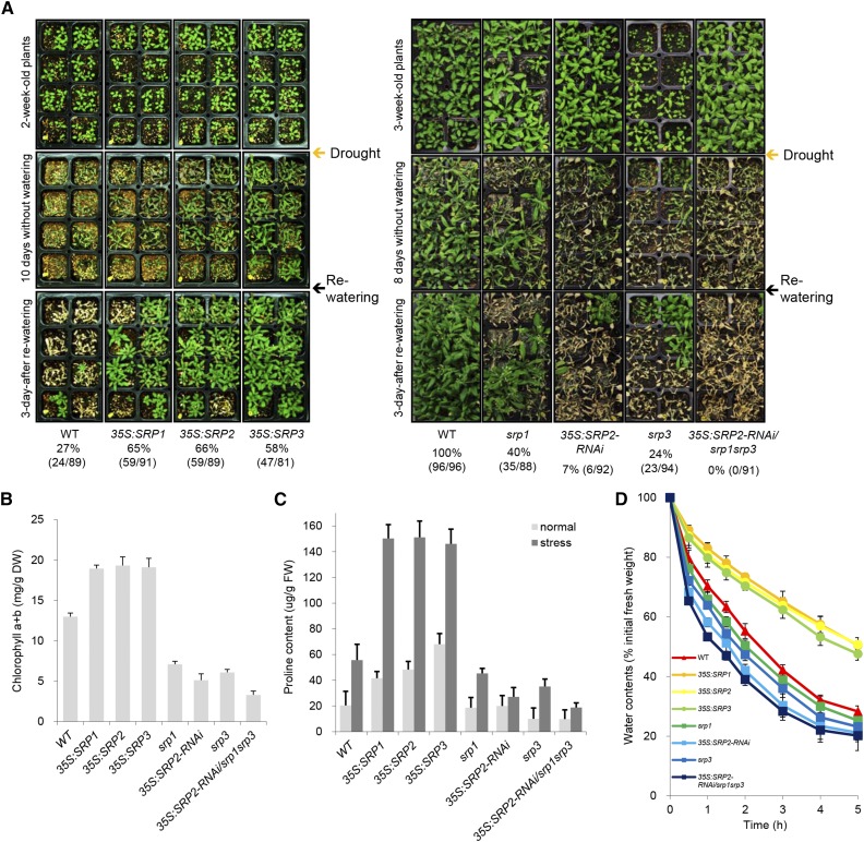
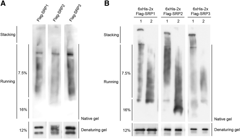
Lipid droplets (LDs) act as repositories for fatty acids and sterols, which are used for various cellular processes such as energy production and membrane and hormone synthesis. LD-associated proteins play important roles in seed development and germination, but their functions in postgermination growth are not well understood. Arabidopsis (Arabidopsis thaliana) contains three SRP homologs (SRP1, SRP2, and SRP3) that share sequence identities with small rubber particle proteins of the rubber tree (Hevea brasiliensis). In this report, the possible cellular roles of SRPs in postgermination growth and the drought tolerance response were investigated. Arabidopsis SRPs appeared to be LD-associated proteins and displayed polymerization properties in vivo and in vitro. SRP-overexpressing transgenic Arabidopsis plants (35S:SRP1, 35S:SRP2, and 35S:SRP3) exhibited higher vegetative and reproductive growth and markedly better tolerance to drought stress than wild-type Arabidopsis. In addition, constitutive over-expression of SRPs resulted in increased numbers of large LDs in postgermination seedlings. In contrast, single (srp1, 35S:SRP2-RNAi, and srp3) and triple (35S:SRP2-RNAi/srp1srp3) loss-of-function mutant lines exhibited the opposite phenotypes. Our results suggest that Arabidopsis SRPs play dual roles as positive factors in postgermination growth and the drought stress tolerance response. The possible relationships between LD-associated proteins and the drought stress response are discussed.


Figures

Figure 1.
Figure 1.
Characterization of Arabidopsis SRPs. A, Schematic structures of SRPs. Solid bars represent the coding regions. Solid lines depict the 5′-and 3′-untranslated regions. Arrows indicate the primers used for genotyping PCR and RT-PCR. Nucleotide sequences of the primers are listed in Supplemental Table S1. B, Subcellular localizations of SRPs in tobacco protoplasts. Protoplasts were obtained from tobacco leaves that transiently expressed 35S:SRP-GFP fusion constructs. The GFP signals were detected by fluorescence microscopy under dark-field or light-field conditions. Scale bars = 10 μm. C, Membrane-association assay of SRPs. The 35S:Flag-SRP fusion genes were transiently expressed in tobacco leaves. Soluble (S) and pellet (P) fractions of total leaf protein extracts were treated with Triton X-100 and subjected to immuno-blot analysis using anti-Flag antibody. D, Lectin-binding assay of SRPs. Total protein extracts were prepared from Flag-SRP-expressing tobacco leaves and were incubated with Con A-sepharose resin in the presence or absence of methyl α-d-glucopyranoside. Proteins eluted from the sepharose resin were analyzed by protein gel-blotting using anti-Flag antibody. Methyl α-d-glucopyranoside was used as a competitor to bind to Con A. KOR1 was used as a positive glycoprotein marker.
Figure 2.
Figure 2.
Expression of SRPs. A, Spatial expression patterns of SRPs in Arabidopsis. Transcript levels of SRP1, SRP2, and SRP3 were analyzed in seedlings, roots, leaves, stems, flowers, siliques, and seeds by real-time qRT-PCR. The expression fold of SRPs was normalized to that of glyceraldehyde-3-phosphate dehydrogenase C subunit, which was used as a reference gene. Results are presented as means ± sd from three independent biological replicates. B, Promoter activities of SRP1, SRP2, and SRP3 in developing Arabidopsis tissues. Histochemical GUS expression profiles in T3 pSRP-GUS transgenic plants were visualized by cyclohexylammonium salt staining for 16 h. Arrow indicates low level of pSRP2-GUS activity in the immature leaves of 7-d-old seedlings. DAF, day after flowering. Scale bars = 0.25 cm. C, qRT-PCR analysis of SRPs. Light-grown, 7-d-old, wild-type Arabidopsis seedlings were subjected to drought (0, 1.5, and 3 h), low temperature (4°C for 0, 1.5, and 3 h), high salinity (300 mm NaCl for 0 and 4.5 h), and 100 μm ABA (0, 1.5, and 3 h). Total RNA was extracted from the treated tissues and analyzed by qRT-PCR. Glyceraldehyde-3-phosphate dehydrogenase C subunit mRNA was used as an internal control for normalization. Error bars indicate means ± sd from three independent experiments.
Figure 3.
Figure 3.
Phenotypic characterization of SRP-overexpressing and loss-of-function srp mutant plants. A, Germination assay. Left: wild-type (WT), SRP-over-expressing, and srp mutant seeds were germinated on half-strength Murashige and Skoog medium. Scale bars = 0.5 cm. Right: germination rates with respect to radicle emergency were determined 1 to 5 d after imbibition. Error bars indicate means ± sd (n = 80). B, Root growth. Left: morphological comparisons of wild-type, SRP-over-expressing, and srp mutant seedlings 4 d after germination. Scale bars = 2.0 cm. Right: growth profiles of wild-type, SRP-over-expressing, and srp mutant early roots 1 to 8 d after germination. Error bars indicate means ± sd (n = 40). C, Leaf growth. Left: morphological comparisons of 2-week-old rosettes from wild-type, SRP-over-expressing, and srp mutant plants. Scale bars = 2.0 cm. Right: quantification of the dimensional parameters of the second leaf. Detached second leaves were analyzed using the SCIONIMAGE program. Error bars indicate means ± sd (n = 40). D, Bolting. Top: bolting phenotypes of wild-type, SRP-over-expressing, and srp mutant plants. Bottom: growth rates of the inflorescence stems of wild-type, SRP-overexpressing, and srp mutant plants were monitored 19 to 31 d after germination. Error bars indicate means ± sd (n = 80).
Figure 4.
Figure 4.
Cellular phenotypes of 35S:SRP transgenic and srp mutant plants. A, Left: longitudinal sections of 3-d-old roots from wild-type, SRP-over-expressing, and srp mutant plants. Vertical lines indicate the PM. Arrows represent the transition zone between the PM and elongation-differentiation zones. The insets show a magnified transition zone. Root sections were stained with propidium iodide and visualized by confocal microscopy. Right: measurement of meristem cell number. Error bars indicate means ± sd (n = 30). B, Different cell sizes in wild-type, SRP-over-expressing, and srp mutant leaves. The epidermal and mesophyll cell layers of 5-d-old leaves were treated with chloral hydrate, a tissue-clearing reagent. The image was visualized by light microscopy. Scale bars = 20 μm.
Figure 5.
Figure 5.
Drought sensitivity of wild-type, SRP-over-expressing, and srp mutant plants. A, Left: Light-grown, 2-week-old wild-type (WT) and SRP over-expressors (35S:SRP1, 35S:SRP2, and 35S:SRP3) were further grown for 10 d without irrigation. Plants were rewatered, and the survival rates were determined 3 d after rewatering. Right: wild-type and srp mutant (srp1, 35S:SRP2-RNAi, srp3, and 35S:SRP2-RNAi/srp1srp3) plants were grown for 3 weeks under normal growth conditions. After 8 d of desiccation, the plants were recovered by rewatering, and the number of surviving plants was counted 3 d after irrigation. B, Total chlorophyll amounts (mg/g DW) in wild-type, SRP-over-expressing, and srp mutant leaves after drought stress. Error bars indicate means ± sd (n = 20). C, Pro content of wild-type, SRP-over-expressing, and srp mutant leaves before (gray bars) and after (dark bars) water stress. Error bars indicate means ± sd (n = 15). D, Measurement of the rate of water loss in cut rosettes from wild-type, SRP-over-expressing, and srp mutant plants. Detached rosette leaves were subjected to desiccation for 0 to 5 h at room temperature. Water loss is represented as the percentage of the initial fresh weight of the detached rosette leaves. Error bars indicate means ± sd (n = 8).
Figure 6.
Figure 6.
Surface structure of seed coats and the expression levels of cell wall-related genes. A, SEM analysis of dry seeds of wild-type (WT), 35S:SRP transgenic, and srp mutant plants. Scale bar = 40 μm. B, Transcriptional changes in cell wall-modifying genes (WAK1, FEI1, and PME3) in SRP-over-expressing and mutant progeny relative to wild-type plants. Data represent means ± sd from three independent experiments.
Figure 7.
Figure 7.
Colocalization of Arabidopsis SRPs to LDs. The 35S:P19 (A), 35S:P19 + 35S:LEC2 (B), and 35S:P19 + 35S:LEC2 + 35S:SRPs-GFP (C) constructs were transiently expressed in tobacco leaves via Agrobacterium-mediated infiltration. P19, a viral suppressor of posttranscriptional gene-silencing, was used to increase infiltration efficiency. LEC2 was used to promote lipid accumulation and increase number of LDs. After 3 d of infiltration, leaves were stained using Nile red (2 μg/mL) and fluorescent signals were visualized via confocal microscopy. Red and green signals represented LDs and SRP-GFPs, respectively. Scale bars = 20 μm.
Figure 8.
Figure 8.
Distribution of LDs and the expression levels of mitochondrial respiratory genes. A, TEM analysis of 6-d-old hypocotyls from wild-type (WT), SRP over-expressors, and 35S:SRP2-RNAi/srp1srp3 triple mutant plants. LDs were divided into six groups (>2.5, 2.0-2.5, 1.5-2.0, 1.0-1.5, 0.5-1.0, and 0.0-0.5 μm) depending on their sizes. Red arrowheads indicate LDs. V, central vacuole; S, starch granule. Scale bars = 10 μm. B, Expression of COX1 and COX2, mitochondrial respiratory marker genes, in wild-type, 35S:SRP transgenic, and 35S:SRP2-RNAi/srp1srp3 mutant plants. Data represent means ± sd from three independent experiments.
Figure 9.
Figure 9.
Polymerization properties of SRPs. A, In vivo polymerization of SRPs. Total protein extracts were prepared from 35S:Flag-SRPs-infiltrated tobacco leaves and subjected to immuno-blot analysis by native (7.5% and 16%; top) or denaturing (12%; bottom) PAGE gel with anti-Flag antibody. B, Lane 1: bacterially expressed 6×His-2×Flag-fused SRPs were purified and subjected to immuno-blot analysis by native (7.5% and 16%; top) or denaturing (12%; bottom) PAGE gel with anti-Flag antibody. Lane 2: 6×His-2×Flag-fused SRPs were boiled in the presence of 0.2% SDS and 100 mm β-mercaptoethanol. Denatured 6×His-2×Flag-SRPs were renatured for 2 h at 28°C and subjected to immuno-blotting by native (top) or denaturing (bottom) PAGE gel with anti-Flag antibody.

Similar articles

Cited by

References

    1. Abe H, Yamaguchi-Shinozaki K, Urao T, Iwasaki T, Hosokawa D, Shinozaki K (1997) Role of Arabidopsis MYC and MYB homologs in drought- and abscisic acid-regulated gene expression. Plant Cell 9: 1859–1868 - PMC - PubMed
    1. Aon MA, Bhatt N, Cortassa SC (2014) Mitochondrial and cellular mechanisms for managing lipid excess. Front Physiol 5: 282. - PMC - PubMed
    1. Atia A, Debez A, Barhoumi Z, Abdelly C, Smaoui A (2009) Histochemical localization of essential oils and bioactive substances in the seed coat of the halophyte Crithmum maritimum L. (Apiaceae). J Plant Biol 52: 448–452
    1. Aubert Y, Vile D, Pervent M, Aldon D, Ranty B, Simonneau T, Vavasseur A, Galaud JP (2010) RD20, a stress-inducible caleosin, participates in stomatal control, transpiration and drought tolerance in Arabidopsis thaliana. Plant Cell Physiol 51: 1975–1987 - PubMed
    1. Bae H, Choi SM, Yang SW, Pai H-S, Kim WT (2009) Suppression of the ER-localized AAA ATPase NgCDC48 inhibits tobacco growth and development. Mol Cells 28: 57–65 - PubMed

Publication types

MeSH terms