. 2016 Mar 8;113(10):2732-7.
doi: 10.1073/pnas.1519044113. Epub 2016 Feb 22.

Rhythmic expression of cryptochrome induces the circadian clock of arrhythmic suprachiasmatic nuclei through arginine vasopressin signaling

Affiliations

Rhythmic expression of cryptochrome induces the circadian clock of arrhythmic suprachiasmatic nuclei through arginine vasopressin signaling

Mathew D Edwards et al. Proc Natl Acad Sci U S A. .

Abstract

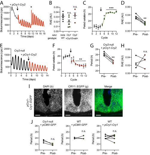
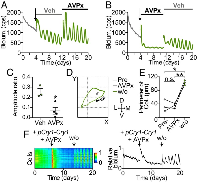
Circadian rhythms in mammals are coordinated by the suprachiasmatic nucleus (SCN). SCN neurons define circadian time using transcriptional/posttranslational feedback loops (TTFL) in which expression of Cryptochrome (Cry) and Period (Per) genes is inhibited by their protein products. Loss of Cry1 and Cry2 stops the SCN clock, whereas individual deletions accelerate and decelerate it, respectively. At the circuit level, neuronal interactions synchronize cellular TTFLs, creating a spatiotemporal wave of gene expression across the SCN that is lost in Cry1/2-deficient SCN. To interrogate the properties of CRY proteins required for circadian function, we expressed CRY in SCN of Cry-deficient mice using adeno-associated virus (AAV). Expression of CRY1::EGFP or CRY2::EGFP under a minimal Cry1 promoter was circadian and rapidly induced PER2-dependent bioluminescence rhythms in previously arrhythmic Cry1/2-deficient SCN, with periods appropriate to each isoform. CRY1::EGFP appropriately lengthened the behavioral period in Cry1-deficient mice. Thus, determination of specific circadian periods reflects properties of the respective proteins, independently of their phase of expression. Phase of CRY1::EGFP expression was critical, however, because constitutive or phase-delayed promoters failed to sustain coherent rhythms. At the circuit level, CRY1::EGFP induced the spatiotemporal wave of PER2 expression in Cry1/2-deficient SCN. This was dependent on the neuropeptide arginine vasopressin (AVP) because it was prevented by pharmacological blockade of AVP receptors. Thus, our genetic complementation assay reveals acute, protein-specific induction of cell-autonomous and network-level circadian rhythmicity in SCN never previously exposed to CRY. Specifically, Cry expression must be circadian and appropriately phased to support rhythms, and AVP receptor signaling is required to impose circuit-level circadian function.

Keywords: arginine vasopressin; bioluminescence; clock; oscillation; period.


Conflict of interest statement

The authors declare no conflict of interest.

Figures

Fig. 1.
Fig. 1.
Induction of molecular pacemaking in arrhythmic Cry1/2-null SCN. (A) EGFP signal from Cry1/2-null SCN transduced with AAV-pCry1-Cry1::EGFP. ITR: inverted terminal repeats; WPRE: WHP posttranscriptional response element. (Scale bar: 10 μm.) (B) PER2::LUC bioluminescence from a Cry1/2-null SCN before (gray) and after (green) transduction (arrow). Asterisk indicates medium change. (C) The period of Cry1- and Cry2- induced rhythms compared with WT (n = 6, 4, 6). All error bars represent mean ± SEM; ****P < 0.0001 vs. WT. (D) Bioluminescence from a Cry1-null SCN before (black) and after (green) transduction with pCry1-Cry1::EGFP. (E) Period of Cry1-null (n = 5) SCN before and after transduction with Cry AAVs; ***P < 0.001 vs. pretreatment, paired t-test. (F) Actogram of Cry1-null mouse before and after stereotaxic injection of Cry1 AAVs into the SCN. (G) The free-running period of Cry1-null animals in continuous darkness before and after Cry1 AAV injection. ***P < 0.001 vs. pretreatment, paired t test (n = 8). (H) Time-lapse images of PER2::LUC bioluminescence from a Cry1/2-null SCN before (Top) and after (Bottom) transduction with pCry1-Cry1::EGFP. CT: circadian time. (Scale bar: 100 μm.) (I) Raster plots of bioluminescence from single cells in H. (J) Normalized PER2::LUC bioluminescence (magenta) and EGFP fluorescence (green/red) over 3 d from Cry1/2-null SCN + pCry1-Cry1::EGFP (Left) or pCry1-Cry2::EGFP (Right). (K) Peak phase of EGFP fluorescence (CT 12 defined as PER2::LUC peak) of CRY1::EGFP and CRY2::EGFP (n = 4, 3).
Fig. S1.
Fig. S1.
Representative confocal images of Cry1/2-null SCN neurons transduced with (A) pCry1-Cry2::EGFP; mean transduction efficiency: 83.2 ± 6.1%. (B) pSyn1-Cry1::EGFP: 86.6 ±1 0.6%. (C) pBmal1-Cry1::EGFP: 79.1 ± 5.6%. n ≥ 3 SCN for each. (Scale bar: 20 μm.)
Fig. S2.
Fig. S2.
(A) Representative Per2::Luc bioluminescence from a Cry1/2-null SCN before (gray) and after (red) transduction with pCry1-Cry2::EGFP (arrow). (B) The RAE of molecular rhythms from WT SCN (n = 6), compared with Cry1/2-null SCN transduced with either pCry1-Cry1::EGFP (n = 6) or pCry1-Cry2::EGFP (n = 4). WT: 0.030 ± 0.003 AU; Cry1-induced: 0.019 ± 0.004 AU; Cry2-induced: 0.030 ± 0.001 AU: P = 0.06; P = 0.75 vs. WT, respectively. (C and D) The peak-to-peak period (C) and RAE (D) (0.042 ± 0.002 AU vs. 0.021 ± 0.001 AU; **P < 0.01) of Cry1-null SCN before and after pCry1-Cry1::EGFP transduction (n = 5). Significance is pretransduction cycle comparison using paired t test (two-tailed). (EH) Representative bioluminescence (E), peak-to-peak period (F), period (G) (26.1 ± 0.3 h vs. 23.2 ± 0.3 h; **P < 0.01) and RAE (H) (0.026 ± 0.013 AU vs. 0.031 ± 0.014 AU; P = 0.65) from Cry2-null SCN before (black) and after (red) transduction with pCry1-Cry2::EGFP (n = 4). (I) Representative image of a Cry1-null SCN transduced in vivo with pCry1-Cry1::EGFP. (Scale bar: 100 μm.) (J) The free-running period of animals before and after stereotaxic injection with control AAVs. Cry1-null + CMV-GFP, pre: 23.2 ± 0.1 h vs. post: 22.9 ± 0.1 h; P = 0.06, WT + CMV-GFP, pre: 24.0 ± 0.1 h vs. post: 24.0 ± 0.1 h, P = 0.65, WT + Cry1, pre: 24.1 ± 0.1 h vs. post: 24.2 ± 0.2 h; P = 0.94.
Fig. S3.
Fig. S3.
(A) Regions of interest, indicative of single oscillators, mapped onto a snapshot of mean bioluminescence over a time-lapse recording of a Cry1/2-null SCN transduced with pCry1-Cry1::EGFP. (B) The period of individual cells from representative WT, Cry1/2-null + pCry1-Cry1::EGFP, and Cry1/2-null + pCry1-Cry2::EGFP SCN. (C) Mean SD of period of single cells in either WT SCN (n = 3 SCN slices; n = 1,230 cells), Cry1/2-null + pCry1-Cry1::EGFP (n = 4 SCN slices; n = 2,146 cells), or pCry1-Cry2::EGFP (n = 3 SCN slices; n = 1,971 cells). (D and E) Rayleigh plots (D) reveal phase coupling of PER::LUC bioluminescence from Cry1- and Cry2-induced rhythms in single cells that were not significantly different from WT SCN as quantified by r mean vector (E). WT: 0.97 ± 0.01; Cry1-induced: 0.91 ± 0.78, P = 0.28 vs. WT; Cry2-induced: 0.90 ± 0.53, P = 0.43 vs. WT.
Fig. 2.
Fig. 2.
Constitutive or phase-delayed expression of Cry1 compromises the SCN molecular clock. (A) Bioluminescence from a Cry1/2-null SCN before (gray) and after (dark green) transduction (arrow) with pSyn1-Cry1::EGFP. Asterisk indicates medium change. (B) Period of molecular rhythms induced by pSyn1-Cry1::EGFP and pBmal1-Cry1::EGFP compared with pCry1-Cry1::EGFP (n = 5, 4, 6; data are from Fig. 1). (C) RAE of induced molecular rhythms. (D) The peak-to-peak period of molecular rhythms in Cry1/2-null SCN induced by different Cry AAVs. Individual SCN represented by different lines. (E) As in A, with pBmal1-Cry1::EGFP (blue). (F) Mean SD of period of single cells in Cry1/2-null SCN transduced with either pSyn1-Cry1::EGFP or pBmal1-Cry1::EGFP (n = 4, 3 slices; n = 660, 1,548 cells) compared with pCry1-Cry1::EGFP. (G) Normalized PER2::LUC bioluminescence (magenta) and EGFP fluorescence (blue) traces over 3 d from SCN transduced with pBmal1-Cry1::EGFP. (H) Mean normalized EGFP fluorescence plotted against normalized PER2::LUC bioluminescence through time for Cry1/2-null SCN transduced with pCry1-Cry1::EGFP or pBmal1-Cry1::EGFP (n = 4, 3). *P < 0.05, **P < 0.01 vs. pCry1. All errors are SEM.
Fig. S4.
Fig. S4.
(A) Representative mCherry fluorescence over 3 d from a WT SCN transduced with an AAV encoding a mCherry fusion protein driven by pSyn1 (n = 3). (B) Representative normalized bioluminescence from NIH 3T3 fibroblast cultures transiently transfected with either pCry1-Luc (green) or pBmal1-Luc (blue). (C) The phase of peak bioluminescence (relative to a peak pPer2-Luc phase of CT12) of pCry1-Luc and pBmal1-Luc (n = 4 for each). pCry1-Luc: CT 15.3 ± 0.3 h; pBmal1-Luc: CT 22.7 ± 0.6 h; ****P < 0.0001. (DG) Representative bioluminescence (D), period (E) [pre: 22.8 ± 02 h vs. post: 23.8 ± 0.6 h; P = 0.24, paired t test (two-tailed)], RAE (F) [pre: 0.027 ± 0.000 AU vs. post: 0.122 ± 0.027 AU; P < 0.05, paired t test (two-tailed)], and peak-to-peak period (G) of Cry1-null SCN before and after (blue) transduction with pBmal1-Cry1::EGFP (arrow; asterisk indicates a medium change) (n = 5). *P < 0.05 vs. pretreatment. (H) Period of individual cells from a representative Cry1/2-null SCN transduced with pCry1-Cry1::EGFP, pSyn1-Cry1::EGFP, or pBmal1-Cry1::EGFP. (I) Representative raster plot of bioluminescence rhythms in single cells in a Cry1/2-null SCN transduced with pBmal1-Cry1::EGFP. (J) Mean normalized EGFP fluorescence plotted against normalized PER2::LUC bioluminescence through time for Cry1/2-null SCN transduced with pCry1-Cry2::EGFP (n = 3).
Fig. 3.
Fig. 3.
Circuit-level organization of molecular rhythms requires appropriately phased, circadian Cry1 expression. (A) Poincaré plots depicting progression of CoL in SCN over three circadian cycles from single SCN, with each cycle represented by a different color. (B) Comparison of the mean perimeter of CoL in different SCN; n = 5, 3, 4, 3, 4, 3 from left to right. *P < 0.05, **P < 0.01 vs. WT, P < 0.05, ††P < 0.01 vs. Cry1/2-null. (C) Poincaré plot depicting CoL over representative cycles in a Cry1/2-null SCN before (gray) and after (green) transduction with pCry1-Cry1::EGFP. (D) Perimeter of CoL over each circadian day before and after transduction (day 0) (n = 4). **P < 0.01, ***P < 0.001 vs. pretransduction mean at cycle−1 (Bonferroni correction).
Fig. 4.
Fig. 4.
AVP signaling is required for Cry-dependent induction of SCN circadian gene expression. (A) Bioluminescence from a Veh-treated (DMSO) Cry1/2-null SCN transduced with pCry1-Cry1::EGFP before application of AVPx. (B) Bioluminescence from a Cry1/2-null SCN transduced with pCry1-Cry1::EGFP in combination with AVPx application before AVPx washout and treatment with vehicle (Veh). (C) Amplitude of induced rhythms under AVPx or Veh, normalized to pretreatment baseline (n = 3, 4). (D) Poincaré plot depicting CoL in Cry1/2-null SCN before (gray) and after (black) transduction with pCry1-Cry1::EGFP and application of AVPx. Subsequent CoL following AVPx washout (w/o) is shown (green). (E) Comparison of the mean perimeter of CoL between conditions in D (n = 3). *P < 0.05, **P < 0.01, paired t tests. (F) Raster plot (Left) and single-cell trace (Right) from SCN in D.
Fig. S5.
Fig. S5.
(A) Representative bioluminescence of a WT SCN before and after AVPx application. (B) Period of WT SCN before, during, and after treatment with either AVPx (n = 5) or Veh (n = 4), increase of 0.5 ± 0.1 h Veh vs. 1.5 ± 0.2 h AVPx; **P < 0.01 vs. Veh. (CE) The change in period (C), amplitude ratio compared with pretreatment (D) (0.37 ± 0.05 Veh vs. 0.48 ± 0.08 AVPx; P = 0.30), and RAE (E) (0.023 ± 0.004 AU Veh vs. 0.023 ± 0.002 AVPx; P = 0.96) of WT SCN rhythms treated with either Veh (gray) or AVPx (black).

References

    1. Hastings MH, Reddy AB, Maywood ES. A clockwork web: Circadian timing in brain and periphery, in health and disease. Nat Rev Neurosci. 2003;4(8):649–661. - PubMed
    1. van der Horst GT, et al. Mammalian Cry1 and Cry2 are essential for maintenance of circadian rhythms. Nature. 1999;398(6728):627–630. - PubMed
    1. Kume K, et al. mCRY1 and mCRY2 are essential components of the negative limb of the circadian clock feedback loop. Cell. 1999;98(2):193–205. - PubMed
    1. Anand SN, et al. Distinct and separable roles for endogenous CRY1 and CRY2 within the circadian molecular clockwork of the suprachiasmatic nucleus, as revealed by the Fbxl3(Afh) mutation. J Neurosci. 2013;33(17):7145–7153. - PMC - PubMed
    1. Ukai-Tadenuma M, et al. Delay in feedback repression by cryptochrome 1 is required for circadian clock function. Cell. 2011;144(2):268–281. - PubMed

Publication types

MeSH terms

LinkOut - more resources