. 2016 Feb 12:7:25.
doi: 10.3389/fphar.2016.00025. eCollection 2016.

Cellular Cholesterol Distribution Influences Proteolytic Release of the LRP-1 Ectodomain

Affiliations

Cellular Cholesterol Distribution Influences Proteolytic Release of the LRP-1 Ectodomain

Bassil Dekky et al. Front Pharmacol. .

Abstract

Low-density lipoprotein receptor-related protein-1 (LRP-1) is a multifunctional matricellular receptor composed of a large ligand-binding subunit (515-kDa α-chain) associated with a short trans-membrane subunit (85-kDa β-chain). LRP-1, which exhibits both endocytosis and cell signaling properties, plays a key role in tumor invasion by regulating the activity of proteinases such as matrix metalloproteinases (MMPs). LRP-1 is shed at the cell surface by proteinases such as membrane-type 1 MMP (MT1-MMP) and a disintegrin and metalloproteinase-12 (ADAM-12). Here, we show by using biophysical, biochemical, and cellular imaging approaches that efficient extraction of cell cholesterol and increased LRP-1 shedding occur in MDA-MB-231 breast cancer cells but not in MDA-MB-435 cells. Our data show that cholesterol is differently distributed in both cell lines; predominantly intracellularly for MDA-MB-231 cells and at the plasma membrane for MDA-MB-435 cells. This study highlights the relationship between the rate and cellular distribution of cholesterol and its impact on LRP-1 shedding modulation. Altogether, our data strongly suggest that the increase of LRP-1 shedding upon cholesterol depletion induces a higher accessibility of the sheddase substrate, i.e., LRP-1, at the cell surface rather than an increase of expression of the enzyme.

Keywords: LRP-1; Raman microspectroscopy; cholesterol; ectodomain; low-density lipoprotein receptor-related protein-1; shedding.


Figures
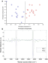
FIGURE 1
FIGURE 1
Cytoplasmic lipid profiling of MDA-MB-231 and MDA-MB-435 cells by Raman analysis. (A) Score plot resulting of the PCA processing of Raman spectra collected on MDA-MB-231 (red circles) and MDA-MB-435 (blue circles) cells. The scores were projected on the two first principal components. (B) Display of the two first principal components. The Raman features comprising the main variability of the spectral data, were highlighted by the grayed underlining.
FIGURE 2
FIGURE 2
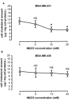
Differential efficiency of MβCD for depleting cholesterol. MDA-MB-231 (A) and MDA-MB-435 (B) breast cancer cells were treated as described under Section “Materials and Methods” with increasing concentrations of MβCD and cellular cholesterol content was then measured. Values expressed as μg cholesterol/mg cell protein are mean ± SD (n = 6 for each cell line). NS, not significant, P < 0.05; Student’s t-test.
FIGURE 3
FIGURE 3
MβCD exhibits different effects on cholesterol depletion in MDA-MB-231 and MDA-MB-435 breast cancer cell lines. MDA-MB-231 (A,B) and MDA-MB-435 (C,D) cells were treated as described under Section “Materials and Methods” with vehicle alone (A,C) or 10 mM MβCD (B,D). Cells were fixed in 3% PFA and treated with filipin (50 μg/mL) for 2 h at room temperature. Projection of each z-stack acquired through confocal microscopy images was merged with DIC images. Filipin labels free cholesterol present in the membranes (red arrow) and in the cytosol (white arrow). Scale bar: 10 μm, n, nucleus.
FIGURE 4
FIGURE 4
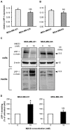
Depletion of cellular cholesterol by MβCD increases shedding of the LRP-1 ectodomain. MDA-MB-231 and MDA-MB-435 cells were treated as described under Section “Materials and Methods” with vehicle alone or 10 mM MβCD. Basal level expression of LRP-1 was measured by quantitative real-time PCR. Data were normalized to ribosomal proteins RPL32 (A) and RS18 (B). Western blotting of LRP-1 α-chain and β-actin was performed from cell lysates, and blotting of LRP-1 α-chain in corresponding amounts of 24-h conditioned medium. Data are from a representative experiment (C). LRP-1 ectodomain shedding was quantified on Western blots of LRP-1 α-chain released in the concentrated conditioned medium (D). NS, not significant, P < 0.05; Student’s t-test.
FIGURE 5
FIGURE 5
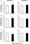
Depletion of cellular cholesterol by MβCD has no effect on the expression of LRP-1 or its sheddases MT1-MMP and ADAM-12. Quantitative real-time PCR of LRP-1 (A), MT1-MMP (B) and ADAM-12 (C) mRNA levels. After normalization to β-actin mRNA amounts (Langlois et al., 2010), data were presented as a percentage of untreated cells. Values are mean ± SD (n = 3 for each cell line). NS, not significant; Student’s t-test.

References

    1. Abramczyk H., Surmacki J., Kopeć M., Olejnik A. K., Lubecka-Pietruszewska K., Fabianowska-Majewska K. (2015). The role of lipid droplets and adipocytes in cancer. Raman imaging of cell cultures: MCF10A, MCF7, and MDA-MB-231 compared to adipocytes in cancerous human breast tissue. Analyst 140 2224–2235. 10.1039/c4an01875c - DOI - PubMed
    1. Baumgart T., Hammond A. T., Sengupta P., Hess S. T., Holowka D. A., Baird B. A., et al. (2007). Large-scale fluid/fluid phase separation of proteins and lipids in giant plasma membrane vesicles. Proc. Natl. Acad. Sci. U.S.A. 104 3165–3170. 10.1073/pnas.0611357104 - DOI - PMC - PubMed
    1. Chang T. Y., Chang C. C. Y., Ohgami N., Yamauchi Y. (2006). Cholesterol sensing, trafficking, and esterification. Annu. Rev. Cell Dev. Biol. 22 129–157. 10.1146/annurev.cellbio.22.010305.104656 - DOI - PubMed
    1. Cremona A., Orsini F., Corsetto P. A., Hoogenboom B. W., Rizzo A. M. (2015). Reversible-dissolution of microdomains in detergent-resistant membranes at physiological temperature. PLoS ONE 10:e0132696 10.1371/journal.pone.0132696 - DOI - PMC - PubMed
    1. Dedieu S., Langlois B., Devy J., Sid B., Schneider C., Henriet P., et al. (2008). LRP-1 silencing prevents malignant cell invasion despite increased pericellular proteolytic activities. Mol. Cell. Biol. 28 2980–2995. 10.1128/MCB.02238-07 - DOI - PMC - PubMed