Review
. 2016 Feb 9:7:30.
doi: 10.3389/fphys.2016.00030. eCollection 2016.

Structural Features of Ion Transport and Allosteric Regulation in Sodium-Calcium Exchanger (NCX) Proteins

Affiliations
Review

Structural Features of Ion Transport and Allosteric Regulation in Sodium-Calcium Exchanger (NCX) Proteins

Moshe Giladi et al. Front Physiol. .

Abstract

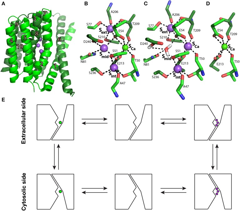
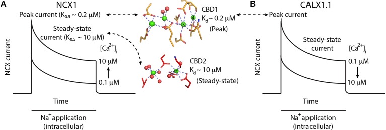
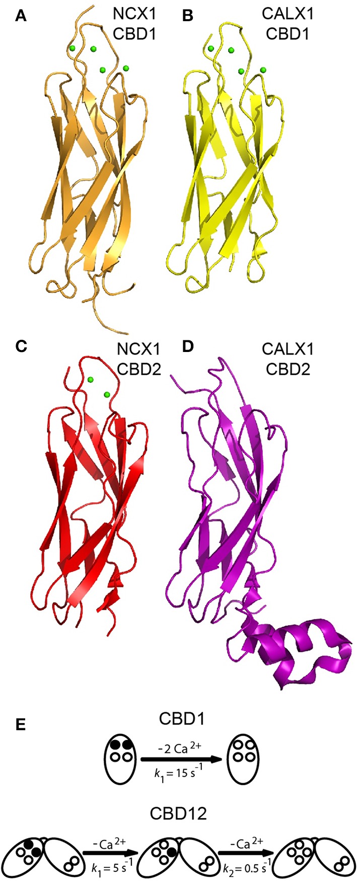
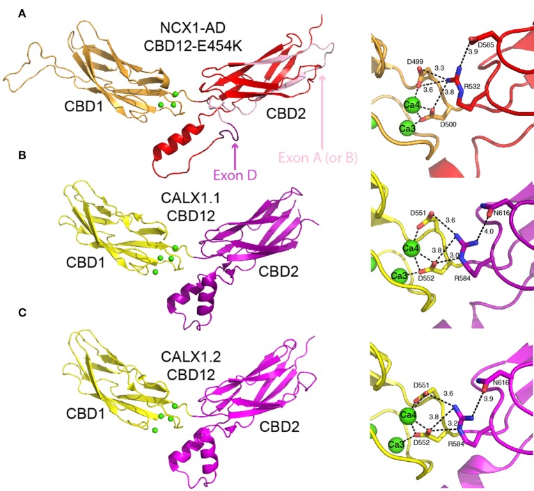
Na(+)/Ca(2+) exchanger (NCX) proteins extrude Ca(2+) from the cell to maintain cellular homeostasis. Since NCX proteins contribute to numerous physiological and pathophysiological events, their pharmacological targeting has been desired for a long time. This intervention remains challenging owing to our poor understanding of the underlying structure-dynamic mechanisms. Recent structural studies have shed light on the structure-function relationships underlying the ion-transport and allosteric regulation of NCX. The crystal structure of an archaeal NCX (NCX_Mj) along with molecular dynamics simulations and ion flux analyses, have assigned the ion binding sites for 3Na(+) and 1Ca(2+), which are being transported in separate steps. In contrast with NCX_Mj, eukaryotic NCXs contain the regulatory Ca(2+)-binding domains, CBD1 and CBD2, which affect the membrane embedded ion-transport domains over a distance of ~80 Å. The Ca(2+)-dependent regulation is ortholog, isoform, and splice-variant dependent to meet physiological requirements, exhibiting either a positive, negative, or no response to regulatory Ca(2+). The crystal structures of the two-domain (CBD12) tandem have revealed a common mechanism involving a Ca(2+)-driven tethering of CBDs in diverse NCX variants. However, dissociation kinetics of occluded Ca(2+) (entrapped at the two-domain interface) depends on the alternative-splicing segment (at CBD2), thereby representing splicing-dependent dynamic coupling of CBDs. The HDX-MS, SAXS, NMR, FRET, equilibrium (45)Ca(2+) binding and stopped-flow techniques provided insights into the dynamic mechanisms of CBDs. Ca(2+) binding to CBD1 results in a population shift, where more constraint conformational states become highly populated without global conformational changes in the alignment of CBDs. This mechanism is common among NCXs. Recent HDX-MS studies have demonstrated that the apo CBD1 and CBD2 are stabilized by interacting with each other, while Ca(2+) binding to CBD1 rigidifies local backbone segments of CBD2, but not of CBD1. The extent and strength of Ca(2+)-dependent rigidification at CBD2 is splice-variant dependent, showing clear correlations with phenotypes of matching NCX variants. Therefore, diverse NCX variants share a common mechanism for the initial decoding of the regulatory signal upon Ca(2+) binding at the interface of CBDs, whereas the allosteric message is shaped by CBD2, the dynamic features of which are dictated by the splicing segment.

Keywords: Ca2+ binding proteins; HDX-MS; NCX; SAXS; X-ray crystallography; allosteric regulation.


Figures

Figure 1
Figure 1
NCX_Mj structure and transport mechanism. (A) Crystal structure of NCX_Mj (PDB 3V5U) in cartoon representation. Helices 1-5 (TM1-5) are light green and helices 6-10 (TM6-10) are dark green. Purple and green spheres represent the Na+ and Ca2+ ions, respectively. (B) Ion coordination, as suggested by the crystal structure of NCX_Mj. (C) 3Na+ ion coordination, as suggested by molecular dynamics simulations and ion-flux assays. (D) Ca2+ binding site. (E) Schematic representation of the ion-exchange mechanism.
Figure 2
Figure 2
Ca2+-dependent regulation of NCX and CALX. (A) Schematic representation of NCX1 currents upon the application of intracellular Na+ to initiate the exchange reaction in the presence of varying [Ca2+]i. Na+ applications initially activates NCX1, followed by a slow decrease in exchange current representing Na+-dependent inactivation. Higher [Ca2+]i results in larger peak currents and reduced Na+-dependent inactivation, as reflected by the higher steady-state current in the presence of 10 μM [Ca2+]i. Binding of Ca2+ ions (green spheres) to the Ca3-Ca4 sites of CBD1 (yellow sticks) results in peak-current activation, whereas the binding of one Ca2+ ion to the CaI site of CBD2 (red sticks) results in steady-state current activation. (B) Schematic representation of CALX1.1 currents upon the application of intracellular Na+ to initiate the exchange reaction in the presence of varying amounts of [Ca2+]i. Na+ application initially activates NCX, followed by a slow decrease in exchange current representing Na+-dependent inactivation. Higher [Ca2+]i results in smaller peak currents and increased Na+-dependent inactivation, as reflected by the smaller steady-state current in the presence of 10 μM [Ca2+]i.
Figure 3
Figure 3
Structures of isolated CBDs and dissociation kinetics. Crystal structures of CBD1 from NCX1 (PDB 2DPK) (A), CBD1 from CALX1 (PDB 3EAD) (B), CBD2 from NCX1-AD (PDB 2QVM) (C), and CBD2 from CALX1.1 (PDB 3E9U) (D) in cartoon representation. (E) Dissociation kinetics of two Ca2+ ions from the Ca3-Ca4 sites of isolated CBD1 and CBD12. The Ca3-Ca4 sites occupied by Ca2+ ions are denoted by filled circles, whereas the open circles represent empty Ca2+ sites. The indicated koff values represent typical values observed in stopped-flow experiments.
Figure 4
Figure 4
Crystal structures of CBD12. Crystal structures of CBD12-E454K from NCX1-AD (PDB 3US9) (A), CBD12 from CALX1.1 (PDB 3RB5) (B), and CBD12 from CALX1.2 (PDB 3RB7) (C) in cartoon representation. Residues participating in the network of interdomain salt bridges are represented as sticks to the right of each structure, with the bond distances within the network indicated. In (A), missing loops were constructed using MODELER. The region corresponding to the mutually exclusive exon is pink, whereas the cassette exon is purple, as indicated by the arrows.
Figure 5
Figure 5
SAXS-EOM analysis and HDX-MS of NCX orthologs and splice variants. Random Rg pools and selected EOM ensemble distributions for CBD12-AD (A), CBD12-BD (B), and Ca2+-bound CALX1.1 and CALX1.2 CBD12 (C). The difference between the HDX profiles of the apo and Ca2+-bound forms of NCX1-CBD12-AD at 10.000 s exchange time (D), CALX1.1-CBD12 at 100 s exchange time (E), and CALX1.2-CBD12 at 100s exchange time (F) are overlaid onto the crystal structures of the different CBD12 proteins (PDB 3US9, 3RB5, and 3RB7, respectively). Ca2+ is indicated as red spheres. The color legend shows the differential HDX after Ca2+ binding.

References

    1. Bernadó P., Mylonas E., Petoukhov M. V., Blackledge M., Svergun D. I. (2007). Structural characterization of flexible proteins using small-angle X-ray scattering. J. Am. Chem. Soc. 129, 5656–5664. 10.1021/ja069124n - DOI - PubMed
    1. Berridge M. J., Bootman M. D., Roderick H. L. (2003). Calcium signalling: dynamics, homeostasis and remodelling. Nat. Rev. Mol. Cell Biol. 4, 517–529. 10.1038/nrm1155 - DOI - PubMed
    1. Bers D. M. (2002). Cardiac excitation-contraction coupling. Nature 415, 198–205. 10.1038/415198a - DOI - PubMed
    1. Besserer G. M., Ottolia M., Nicoll D. A., Chaptal V., Cascio D., Philipson K. D., et al. . (2007). The second Ca2+-binding domain of the Na+ Ca2+ exchanger is essential for regulation: crystal structures and mutational analysis. Proc. Natl. Acad. Sci. U.S.A. 104, 18467–18472. 10.1073/pnas.0707417104 - DOI - PMC - PubMed
    1. Blanchet C. E., Svergun D. I. (2013). Small-angle X-ray scattering on biological macromolecules and nanocomposites in solution. Annu. Rev. Phys. Chem. 64, 37–54. 10.1146/annurev-physchem-040412-110132 - DOI - PubMed

LinkOut - more resources