Review
. 2016 Feb 8:7:35.
doi: 10.3389/fimmu.2016.00035. eCollection 2016.

Clearance Deficiency and Cell Death Pathways: A Model for the Pathogenesis of SLE

Affiliations
Review

Clearance Deficiency and Cell Death Pathways: A Model for the Pathogenesis of SLE

Aparna Mahajan et al. Front Immunol. .

Abstract

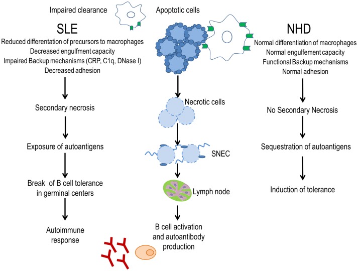
Alterations of cell death pathways, including apoptosis and the neutrophil specific kind of death called NETosis, can represent a potential source of autoantigens. Defects in the clearance of apoptotic cells may be responsible for the initiation of systemic autoimmunity in several chronic inflammatory diseases, including systemic lupus erythematosus (SLE). Autoantigens are released mainly from secondary necrotic cells because of a defective clearance of apoptotic cells or an inefficient degradation of DNA-containing neutrophil extracellular traps (NETs). These modified autoantigens are presented by follicular dendritic cells to autoreactive B cells in germinal centers of secondary lymphoid organs. This results in the loss of self-tolerance and production of autoantibodies, a unifying feature of SLE. Immune complexes (IC) are formed from autoantibodies bound to uncleared cellular debris in blood or tissues. Clearance of IC by blood phagocytes, macrophages, and dendritic cells leads to proinflammatory cytokine secretion. In particular, plasmacytoid dendritic cells produce high amounts of interferon-α upon IC uptake, thereby contributing to the interferon signature of patients with SLE. The clearance of antinuclear IC via Fc-gamma receptors is considered a central event in amplifying inflammatory immune responses in SLE. Along with this, the accumulation of cell remnants represents an initiating event of the etiology, while the subsequent generation of autoantibodies against nuclear antigens (including NETs) results in the perpetuation of inflammation and tissue damage in patients with SLE. Here, we discuss the implications of defective clearance of apoptotic cells and NETs in the development of clinical manifestations in SLE.

Keywords: NETosis; SLE; apoptosis; autoantibody; clearance.


Figures

Figure 1
Figure 1
Evidence for a clearance deficiency in the etiology of SLE. In normal healthy donors (NHD), apoptotic cells are efficiently cleared by macrophages and opsonins, avoiding the exposure of autoantigens and promoting tolerance. In patients with SLE, impaired clearance of dead and dying cells due to less efficient macrophages, deficiency of opsonins, and presence of anti-opsonin antibodies leads to autoantigen exposure through secondary necrotic cells (SNECs) formation. Accumulation of SNECs in germinal centers facilitates autoantigen presentation by follicular dendritic cells to autoreactive B cells. In consequence, the immunological tolerance is compromised and the autoimmune response is initiated.
Figure 2
Figure 2
Model of the pathogenesis of SLE. Impaired clearance of apoptotic cells and NETs is the main contributing factor in the etiopathogenesis of SLE. Inefficient clearance of apoptotic cells leads to the accumulation of secondary necrotic cells (SNECs), along with the release of proinflammatory cytokines by pathologically activated phagocytes. Circulating SNECs are sensitized by autoantibodies. This results in tissues deposition of immune complexes (IC). IC are then cleared by blood-borne phagocytes, such as macrophages and dendritic cells, which consequently release IFN-α and other inflammatory cytokines. The unabated production of IFN-α precipitates cell death and organ damage. Uncleared NETs serve as a source of autoantigen and initiate antineutrophil cytoplasmic antibodies. Internalization of antineutrophil cytoplasmic antibody (ANCA) immune complexes by pDCs causes an enhanced release of IFN-α. Increased IFN-α levels prime neutrophils to undergo NETosis. The presence of DNase-inhibitors, anti-dsDNA autoantibodies, and low levels of opsonins in patients with SLE worsen the clearance of NETs. A vicious cycle is initiated leading to the formation and deposition of more IC, inflammation, cell death, and organ damage. Abbreviations: ANCA, antineutrophil cytoplasmic antibody; IC, immune complex(es); SLE, systemic lupus erythematosus; SNEC, secondary necrotic cell-derived material; NETs, neutrophil extracellular traps; Ag, antigen.

Similar articles

Cited by

References

    1. Galluzzi L, Vitale I, Abrams JM, Alnemri ES, Baehrecke EH, Blagosklonny MV, et al. Molecular definitions of cell death subroutines: recommendations of the Nomenclature Committee on Cell Death 2012. Cell Death Differ (2012) 19(1):107–20.10.1038/cdd.2011.96 - DOI - PMC - PubMed
    1. Kerr JF, Wyllie AH, Currie AR. Apoptosis: a basic biological phenomenon with wide-ranging implications in tissue kinetics. Br J Cancer (1972) 26(4):239–57.10.1038/bjc.1972.33 - DOI - PMC - PubMed
    1. Biermann M, Maueroder C, Brauner JM, Chaurio R, Janko C, Herrmann M, et al. Surface code – biophysical signals for apoptotic cell clearance. Phys Biol (2013) 10(6):065007.10.1088/1478-3975/10/6/065007 - DOI - PubMed
    1. Zirngibl M, Furnrohr BG, Janko C, Munoz LE, Voll RE, Gregory CD, et al. Loading of nuclear autoantigens prototypically recognized by SLE sera into late apoptotic vesicles requires intact microtubules and MLCK activity. Clin Exp Immunol (2015) 179(1):39–49.10.1111/cei.12342 - DOI - PMC - PubMed
    1. Elmore S. Apoptosis: a review of programmed cell death. Toxicol Pathol (2007) 35(4):495–516.10.1080/01926230701320337 - DOI - PMC - PubMed