. 2016:2016:5796305.
doi: 10.1155/2016/5796305. Epub 2016 Jan 21.

Chemokine (C-X-C) Ligand 12 Facilitates Trafficking of Donor Spermatogonial Stem Cells

Affiliations

Chemokine (C-X-C) Ligand 12 Facilitates Trafficking of Donor Spermatogonial Stem Cells

Zhiyv Niu et al. Stem Cells Int. 2016.

Abstract

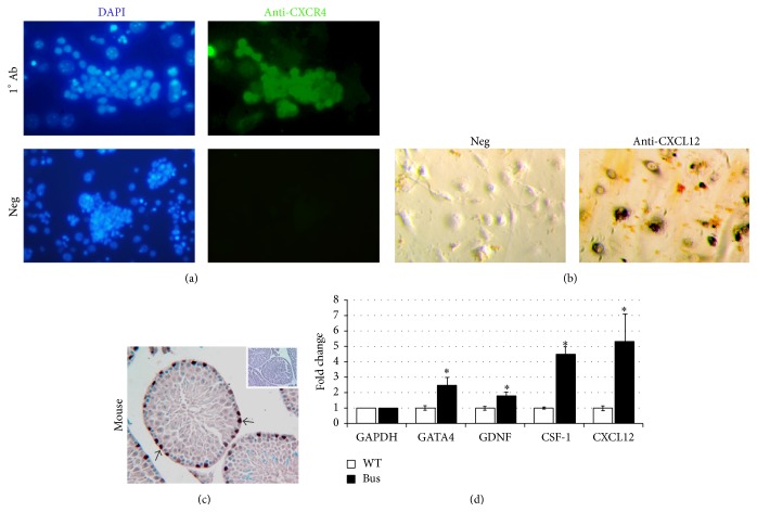
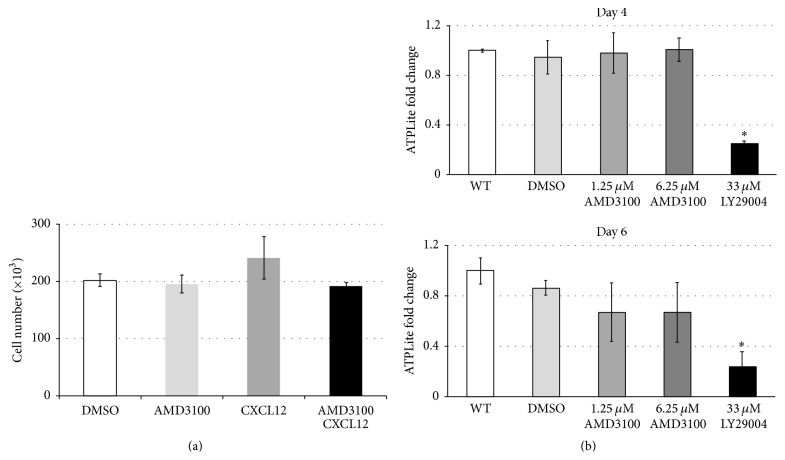
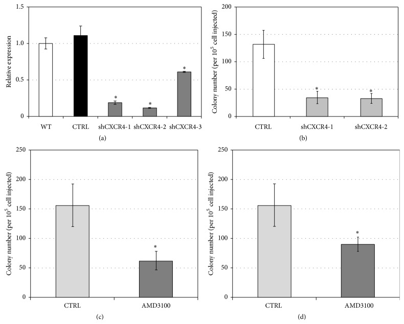
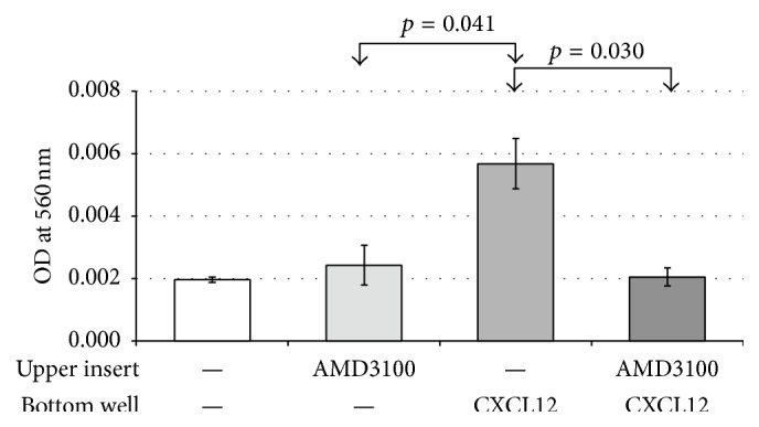
The chemokine (C-X-C) receptor type 4 (CXCR4) is an early marker of primordial germ cells (PGCs) essential for their migration and colonization of the gonads. In spermatogonial stem cells (SSCs), the expression of CXCR4 is promoted by the self-renewal factor, glial cell line-derived neurotrophic factor (GDNF). Here, we demonstrate an important role of CXCR4 during donor mouse SSCs reoccupation of the endogenous niche in recipient testis. Silencing of CXCR4 expression in mouse SSCs dramatically reduced the number of donor stem cell-derived colonies, whereas colony morphology and spermatogenesis were comparable to controls. Inhibition of CXCR4 signaling using a small molecule inhibitor (AMD3100) during the critical window of homing also significantly lowered the efficiency of donor-derived SSCs to establish spermatogenic colonies in recipient mice; however, the self-renewal of SSCs was not affected by exposure to AMD3100. Rather, in vitro migration assays demonstrate the influence of CXCR4-CXCL12 signaling in promoting germ cell migration. Together, these studies suggest that CXCR4-CXCL12 signaling functions to promote homing of SSCs towards the stem cell niche and plays a critical role in reestablishing spermatogenesis.


Figures

Figure 1
Figure 1
In vitro and in vivo expression analysis of CXCR4/CXCL12 in rodent testis cells. (a) Expression of CXCR4 membrane receptor in SSC-enriched cultured germ cells was determined by immunofluorescence using FITC-labeled anti-CXCR4 antibody (green), with the cell nucleus labeled by DAPI (blue). (b) Immunohistochemistry staining of CXCL12 protein in the STO feeder cells. (c) Immunohistochemistry staining of CXCR4 in mouse testis. Arrows show CXCR4 staining of germ cells on the basement membrane of seminiferous tubule. (d) The fold change in gene expression of GATA4, GDNF, CSF-1, and CXCL12 in the testes of untreated and Busulfan-treated recipient mice was verified by qRT-PCR. Changes in gene expression are normalized to GAPDH. Significance (denoted as ) was calculated using Student's t-test (with p < 0.05, n = 3). Data are means ± SEM.
Figure 2
Figure 2
Effects of CXCR4 inhibitor AMD3100 on SSC-enriched THY1+ mouse germ cells. (a) An equal number of SSC-enriched cultured germ cells (1 × 10⁵ cells/well) were cultured on a layer of STO feeder cells and were treated with the vehicle (DMSO) or treated with AMD3100 (1.25 μM), CXCL12 (10 ng/mL), or the combination of both CXCL12 and AMD3100. After 7 days the total number of cultured germ cells was quantified by cell counting. Values are representative of three independent experiments, and significance was calculated using one-way ANOVA. Data are means ± SEM. (b) Impact of AMD3100 on germ cell viability. SSC-enriched cultured germ cells were seeded on feeder-free, laminin-coated 96-well plates at a density of 1 × 10⁴ cells/well. Cells were either untreated (WT) or treated with DMSO control, AMD3100 (1.25 μM and 6.25 μM), or AKT inhibitor, LY29004 (33 μM). Cell viability was assessed at days 4 and 6 following treatment using the ATPLite assay. Data are means ± SEM, where significance (denoted as ) was calculated using Student's t-test (p < 0.05, n = 4).
Figure 3
Figure 3
Inhibition of CXCR4 expression or function reduces stem cell activity of SSC-enriched germ cells. (a) qRT-PCR examination of shRNA-mediated knockdown of CXCR4 gene expression. SSC-enriched germ cell cultures were transduced with lentivirus containing a scrambled control shRNA vector or with one of three CXCR4-targeting shRNA clones (shCXCR4-1, shCXCR4-2, and shCXCR4-3). The transcript levels of CXCR4 were normalized to GAPDH gene expression and compared to untreated controls using 2−ΔΔCT. Significance was calculated using a Student's t-test (p < 0.05; n = 3). Data are means ± SEM. (b) Colony forming efficiency of CXCR4-deficient SSC-enriched germ cell cultures. CXCR4 shRNA lentiviral transduced cultures of SSC-enriched germ cells derived from Rosa-lacZ mice were transplanted at 1 × 10⁴ cells/testis into the testes of Busulfan-treated recipient mice. Donor-derived spermatogenesis was quantified by staining recipient testes with X-gal 2 months after transplantation. Significance was calculated using a Student's t-test (p < 0.05; n = 6 testes per treatment group). Data are means ± SEM. (c) AMD3100 disrupts SSC colony formation. Donor Rosa-lacZ SSC-enriched germ cells were treated with AMD3100 (5 μg/testis) followed by transplantation into recipient testis (data are means ± SEM, p = 0.022, n = 4 testes per treatment group). (d) Alternatively, recipient mice bearing Rosa-lacZ SSC-enriched transplanted germ cells were repeatedly administered AMD3100 (5 μg/testis) via the intraperitoneal (IP) route at 12 hr intervals for 2 days following transplantation. Donor-derived colonies were detected using X-gal and counted 2 months after transplantation. Data are means ± SEM, where significance was assessed using Student's t-test (p = 0.058, n = 2 testes per treatment group).
Figure 4
Figure 4
CXCL12/CXCR4 pathway promotes germ cell migration. SSC-enriched germ cells seeded in upper transwell in the presence or absence of AMD3100 (1.25 μM) were exposed to CXCL12 (10 ng/mL) in the lower well. Measurement of the absorbance (562 nm) in the bottom well was used to assess the active number of germ cells that migrated from upper to lower wells. Data are means ± SEM, where significance was assessed using a Student's t-test (p < 0.05, n = 3 experimental replicates).

References

    1. Brinster R. L. Male germline stem cells: from mice to men. Science. 2007;316(5823):404–405. doi: 10.1126/science.1137741. - DOI - PMC - PubMed
    1. Oatley J. M., Brinster R. L. The germline stem cell niche unit in mammalian testes. Physiological Reviews. 2012;92(2):577–595. doi: 10.1152/physrev.00025.2011. - DOI - PMC - PubMed
    1. Yang Q.-E., Oatley J. M. Spermatogonial stem cell functions in physiological and pathological conditions. Current Topics in Developmental Biology. 2014;107:235–267. doi: 10.1016/b978-0-12-416022-4.00009-3. - DOI - PubMed
    1. Griswold M. D., Oatley J. M. Concise review: defining characteristics of mammalian spermatogenic stem cells. Stem Cells. 2013;31(1):8–11. doi: 10.1002/stem.1253. - DOI - PMC - PubMed
    1. González-González E., López-Casas P. P., Del Mazo J. Gene silencing by RNAi in mouse Sertoli cells. Reproductive Biology and Endocrinology. 2008;6, article 29 doi: 10.1186/1477-7827-6-29. - DOI - PMC - PubMed

LinkOut - more resources