

Rare benign mixed tumour of the upper lip: A case report
- PMID: 26904190
- PMCID: PMC4720717
- DOI: 10.1016/j.amsu.2015.10.001
Rare benign mixed tumour of the upper lip: A case report
Erratum in
-
Erratum to "Rare benign mixed tumour of the upper lip: A case report" [Ann. Med. Surg. 4 (2015) 380-383].Ann Med Surg (Lond). 2016 May 30;11:66. doi: 10.1016/j.amsu.2016.05.015. eCollection 2016 Nov. Ann Med Surg (Lond). 2016. PMID: 29225821 Free PMC article.
Abstract
Background: Chondroid syringomas (CS) are rare benign mixed tumours. Clinical differentiation can be misleading due to the silent presentation, with only histopathological findings confirming the diagnosis.
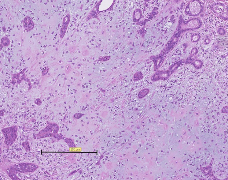
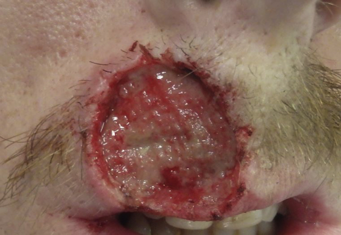
Case report: A 23-year-old Caucasian gentleman presented with an eighteen month history of increasing size of his exophytic upper lip mass. The initial clinical impression was thought to be related to the skin. Following a punch biopsy, histopathology confirmed appearance in keeping with part of a chondroid syringoma with subsequent excision of the lesion.
Discussion: CS present as a slow-growing, asymptomatic, non-tender, nonulcerated, smooth, firm subcutaneous, or intradermal nodule and can range from 0.5 to 3.0 cm, predominantly occurring in the head and neck region in patients aged above 35 years with a male predication. The most effective diagnostic method is microscopic examination. The gold standard treatment modality is by complete excision with a margin of normal tissue in order to examine the histopathologic features and prevent recurrence.
Conclusion: CS should be included as a differential diagnosis of facial subcutaneous skin lesions in middle aged male patients. Careful evaluation, with a view of total excision and adequate surgical margin will enable diagnostic confirmation, whilst maintaining the aesthetic and functional unit.
Keywords: Benign; Chondroid syringoma; Mixed tumour; Upper lip.
Figures


References
-
- Paik Y.S., Liess B.D. Chondroid syringoma of the scalp: case report and discussion of clinical features, histopathology, and treatment. Ear Nose Throat J. 2011;90(4):190–191. - PubMed
-
- Al Omran Y., Mohamed R., Al-Omran M.K. Chondroid syringoma of the ear lobule: a unique case. Ear Nose Throat J. 2014;93(4–5):E62–E63. - PubMed
-
- Chen A.H., Moreano E.H., Houston B., Funk G.F. Chondroid syringoma of the head and neck: clinical management and literature review. Ear Nose Throat J. 1996;72(2):104–108. - PubMed
-
- Triantafyllou A.G., Rapidis A.D. Chondroid syringoma of the upper lip: report of case. J. Oral Maxillfoac. Surg. 1986;44(9):744–748. - PubMed
Publication types
LinkOut - more resources
Full Text Sources
Other Literature Sources
